 Add to Chrome
 Add to Firefox
 Add to Edge
Improving PET/CT-Based Whole-Body Lesion Segmentation Using Prediction Uncertainty-Augmented Models
Jun 08, 2026Accurate lesion segmentation from whole-body Positron Emission Tomography (PET)/Computed Tomography (CT) scans is essential for cancer staging and treatment planning. PET provides functional metabolic information with different radiotracers, while CT offers anatomical localization. Lesion delineation from PET/CT imaging is clinically challenging due to subtle imaging features, confounders, and inter-reader variability. Existing deep learning approaches suffer from training-related stochasticity, inconsistent predictions, missed lesions in high tumor-burden cases, and lack uncertainty quantification, limiting their clinical reliability. Using nnU-Net as a baseline, we propose an uncertainty-aware framework for whole-body PET/CT lesion segmentation that integrates (1) Bayesian ensembling to reduce training stochasticity, (2) voxel-wise uncertainty quantification with epistemic and aleatoric decomposition, and (3) epistemic uncertainty-augmented training to improve lesion detection. Two public datasets, AutoPET-III (1,611 scans) and Deep-PSMA (200 scans), comprising FDG and PSMA studies across multiple cancer types, are used for training and evaluation. Bayesian ensembling improves robustness and performance over deterministic nnU-Net models on the unseen AutoPET-III test set. Uncertainty maps highlight regions of model disagreement and correlate with misclassifications, particularly false positives. Uncertainty-augmented training improves lesion recovery at the cost of increased FPVol, reflecting a precision-recall trade-off. A case-adaptive routing strategy further improves Dice by selecting between the base and augmented models. To our knowledge, this is the first study to systematically investigate uncertainty quantification in multi-tracer, pan-cancer PET/CT segmentation and to combine Bayesian ensembling with uncertainty-aware modeling for this task.
Automatic Post-Stroke Lesion Segmentation on MR Images using 3D Residual Convolutional Neural Network
Nov 25, 2019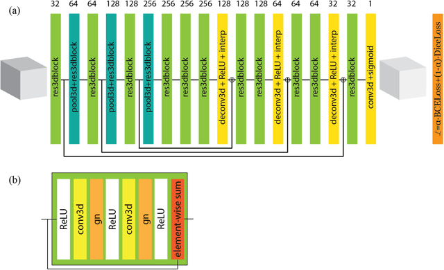
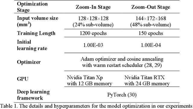
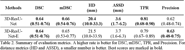

In this paper, we demonstrate the feasibility and performance of deep residual neural networks for volumetric segmentation of irreversibly damaged brain tissue lesions on T1-weighted MRI scans for chronic stroke patients. A total of 239 T1-weighted MRI scans of chronic ischemic stroke patients from a public dataset were retrospectively analyzed by 3D deep convolutional segmentation models with residual learning, using a novel zoom-in&out strategy. Dice similarity coefficient (DSC), Average symmetric surface distance (ASSD), and Hausdorff distance (HD) of the identified lesions were measured by using the manual tracing of lesions as the reference standard. Bootstrapping was employed for all metrics to estimate 95% confidence intervals. The models were assessed on the test set of 31 scans. The average DSC was 0.64 (0.51-0.76) with a median of 0.78. ASSD and HD were 3.6 mm (1.7-6.2 mm) and 20.4 mm (10.0-33.3 mm), respectively. To the best of our knowledge, this performance is the highest achieved on this public dataset. The latest deep learning architecture and techniques were applied for 3D segmentation on MRI scans and demonstrated to be effective for volumetric segmentation of chronic ischemic stroke lesions.