 Add to Chrome
 Add to Firefox
 Add to Edge
FetSelect: Task-Specific Architectures and Self-Supervised Learning for Automated Fetal Ultrasound Frame Selection
Jun 21, 2026
Automated frame selection for fetal biometry remains under addressed, with most prior work targeting generic quality assessment or downstream measurement pipelines that assume suitable frames are available. We introduce FetSelect, a task-specific framework that pairs a frozen vision foundation backbone with a hybrid multi-head design: a Task-Gated classification head and a Detection-derived quality head combined via learned fusion. We curate 6,486 expert-labeled frames across four targets: Crown-Rump Length (CRL), Nuchal Translucency (NT), Nasal Bone (NB), and Scalebar, and adapt the backbone with BYOL pretraining on 19,019 unlabeled images. On a held-out test set (974 frames), FetSelect achieves mean AUROC 0.956 and mean correlation 0.818 with expert quality annotations. Ablations confirm that hybrid fusion surpasses single-head variants, and ultrasound-specific self-supervision yields consistent gains. Evaluation on external clinical videos and 509 external CRL images demonstrates task-specific discrimination.

FADA: Accessible fetal ultrasound interpretation and annotation with a selectively distilled unified vision-language model
Jun 09, 2026A global shortage of trained sonographers limits prenatal ultrasound screening in low- and middle-income countries, where over half of pregnant women receive no skilled sonography. Current deep learning approaches address detection, segmentation, or classification in isolation, each demanding a separate model and expert-specified labels at inference. We present FADA, a unified vision-language model built on Qwen3.5-VL that performs clinical interpretation, classification, detection, and segmentation through a single interpretation-first pipeline without external labels. FADA distills knowledge from four domain-specific foundation models (FetalCLIP, UltraSAM, USF-MAE, UltraFedFM) via offline pre-computed feature caching. Selective distillation, which applies feature alignment only to annotation tasks while interpretation relies on standard fine-tuning, consistently outperforms full distillation across most evaluation axes. The recommended variant, FADA-SKD, achieves 0.8820 mean Dice for segmentation, 0.7671 mAP@0.50 for detection, and 100% structured interpretation compliance. Expert sonographer validation across 237 images confirms clinically acceptable outputs in both autonomous and human-in-the-loop modes, with 73.5% of interpretations scoring perfectly under clinician guidance. The system is trainable on a single consumer GPU and deployable without cloud connectivity. We validate edge deployment by running the compressed 0.8B model on a commodity smartphone (Qualcomm Snapdragon 7 Gen 1, 12 GB RAM) using llama.cpp with GGUF quantization, completing the full 5-phase pipeline in approximately 60 seconds entirely offline. This establishes a practical pathway for integrating AI-assisted fetal assessment with portable ultrasound devices, directly addressing diagnostic access gaps in resource-constrained settings. Code, models, and data are available at https://github.com/mahmoodphd/FADA.
Focus on What Matters: Two-Stage ROI-Aware Refinement for Anatomy-Preserving Fetal Ultrasound Reconstruction
Apr 26, 2026Measurement-critical ultrasound tasks often depend on a small anatomical region, making global reconstruction metrics an unreliable proxy for clinical fidelity. We propose an ROI-aware representation learning framework and instantiate it for first-trimester nuchal translucency (NT) screening under multi-hospital domain shift. A two-phase convolutional autoencoder (CAE) first learns a globally faithful 128-D latent code via MS-SSIM, then refines the NT ROI using intensity (L1) and normalized Sobel-edge constraints. To combine these heterogeneous objectives without manual tuning, we initialize loss weights via gradient-based calibration from per-term gradient magnitudes. Under strict hospital-wise evaluation with one hospital held out, ROI refinement improves both global and measurement-relevant quality: on the standard dev split it increases PSNR by +0.27 dB (val) and +0.29 dB (held-out test), reduces ROI MAE by 8.87% (val) and 6.43% (held-out test), and reduces ROI Edge-MAE by 11.10% on source hospitals and 4.90% on the unseen hospital. Beyond reconstruction, frozen-latent probes provide additional evidence of generalization: hospital provenance becomes less confidently predictable on the unseen site (0.556 to 0.541 max-softmax; 0.684 to 0.688 entropy) while OOD detection remains strong across site-held-out protocols (Mahalanobis AUROC up to 0.9956, with modest KNN gains in challenging splits). The same ROI-aware refinement principle is anatomy-agnostic and can be adopted for other fetal biometry targets (e.g., crown-rump length (CRL), nasal bone (NB)) and broader medical imaging settings where small ROIs dominate clinical decisions.
Multi-Modal Perceiver Language Model for Outcome Prediction in Emergency Department
Apr 03, 2023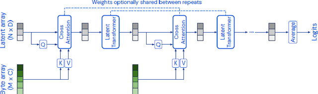

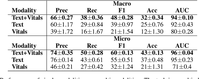
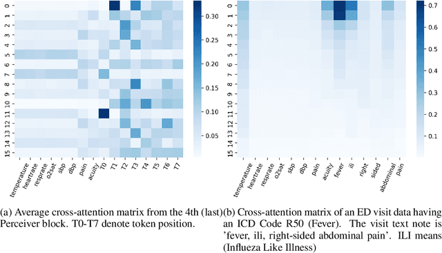
Language modeling have shown impressive progress in generating compelling text with good accuracy and high semantic coherence. An interesting research direction is to augment these powerful models for specific applications using contextual information. In this work, we explore multi-modal language modeling for healthcare applications. We are interested in outcome prediction and patient triage in hospital emergency department based on text information in chief complaints and vital signs recorded at triage. We adapt Perceiver - a modality-agnostic transformer-based model that has shown promising results in several applications. Since vital-sign modality is represented in tabular format, we modified Perceiver position encoding to ensure permutation invariance. We evaluated the multi-modal language model for the task of diagnosis code prediction using MIMIC-IV ED dataset on 120K visits. In the experimental analysis, we show that mutli-modality improves the prediction performance compared with models trained solely on text or vital signs. We identified disease categories for which multi-modality leads to performance improvement and show that for these categories, vital signs have added predictive power. By analyzing the cross-attention layer, we show how multi-modality contributes to model predictions. This work gives interesting insights on the development of multi-modal language models for healthcare applications.
Towards deep observation: A systematic survey on artificial intelligence techniques to monitor fetus via Ultrasound Images
Jan 17, 2022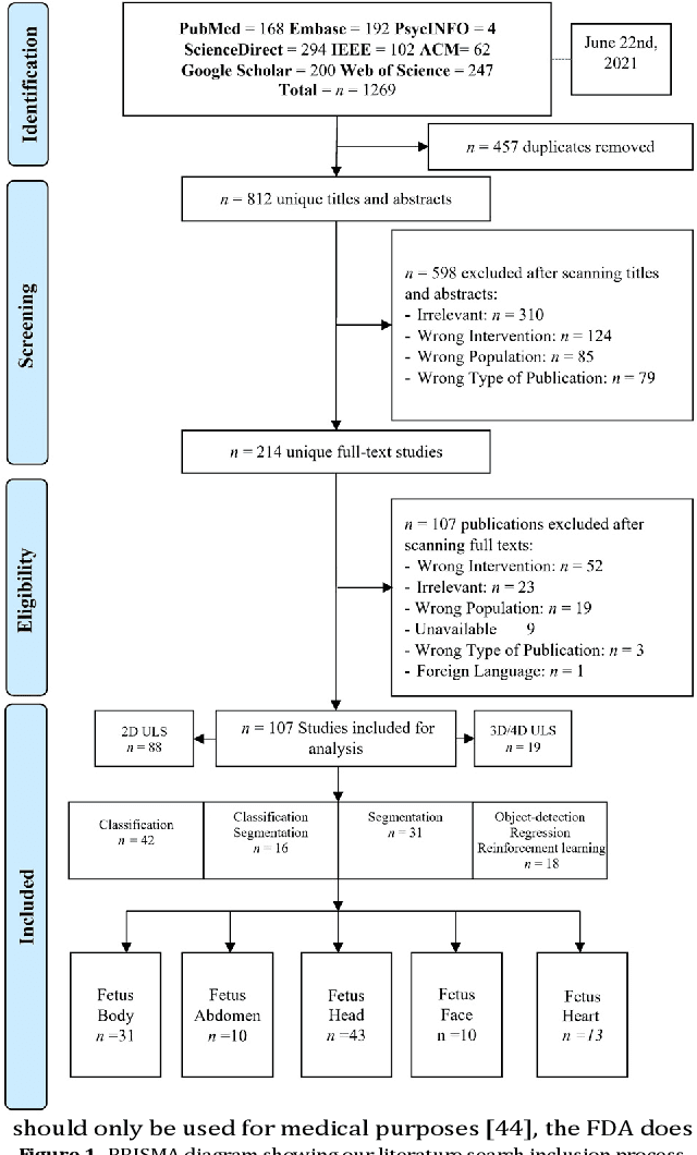
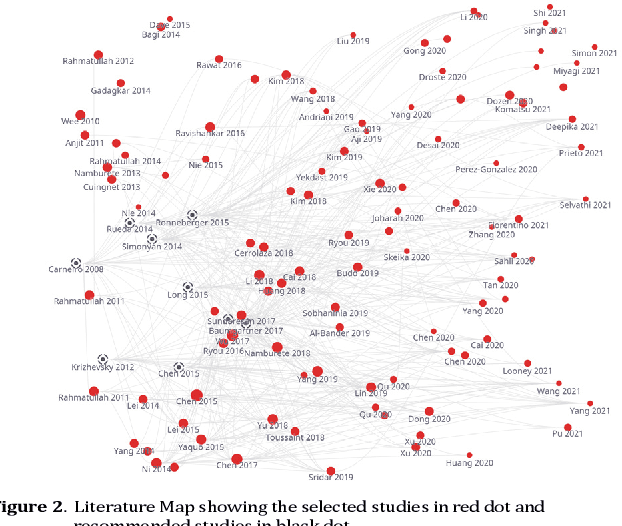
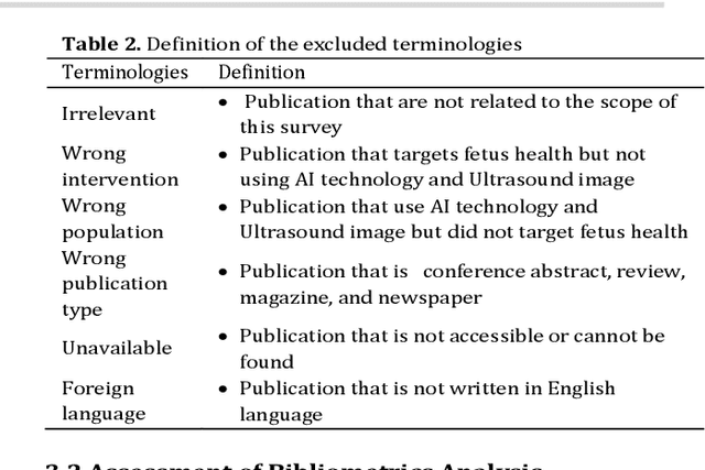
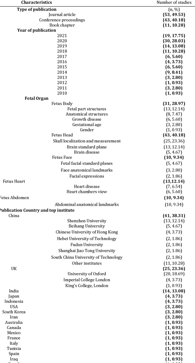
Developing innovative informatics approaches aimed to enhance fetal monitoring is a burgeoning field of study in reproductive medicine. Several reviews have been conducted regarding Artificial intelligence (AI) techniques to improve pregnancy outcomes. They are limited by focusing on specific data such as mother's care during pregnancy. This systematic survey aims to explore how artificial intelligence (AI) can assist with fetal growth monitoring via Ultrasound (US) image. We used eight medical and computer science bibliographic databases, including PubMed, Embase, PsycINFO, ScienceDirect, IEEE explore, ACM Library, Google Scholar, and the Web of Science. We retrieved studies published between 2010 to 2021. Data extracted from studies were synthesized using a narrative approach. Out of 1269 retrieved studies, we included 107 distinct studies from queries that were relevant to the topic in the survey. We found that 2D ultrasound images were more popular (n=88) than 3D and 4D ultrasound images (n=19). Classification is the most used method (n=42), followed by segmentation (n=31), classification integrated with segmentation (n=16) and other miscellaneous such as object-detection, regression and reinforcement learning (n=18). The most common areas within the pregnancy domain were the fetus head (n=43), then fetus body (n=31), fetus heart (n=13), fetus abdomen (n=10), and lastly the fetus face (n=10). In the most recent studies, deep learning techniques were primarily used (n=81), followed by machine learning (n=16), artificial neural network (n=7), and reinforcement learning (n=2). AI techniques played a crucial role in predicting fetal diseases and identifying fetus anatomy structures during pregnancy. More research is required to validate this technology from a physician's perspective, such as pilot studies and randomized controlled trials on AI and its applications in a hospital setting.