 Add to Chrome
 Add to Firefox
 Add to Edge
Imaging dynamics beneath turbid media via parallelized single-photon detection
Jul 22, 2021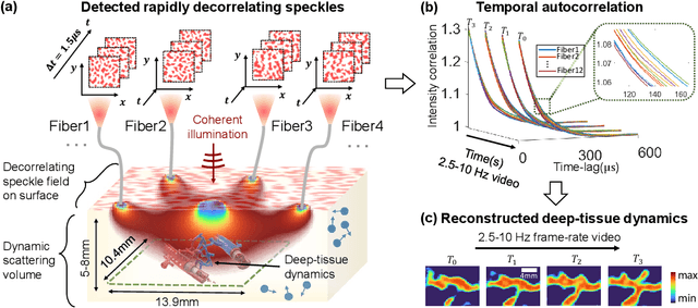
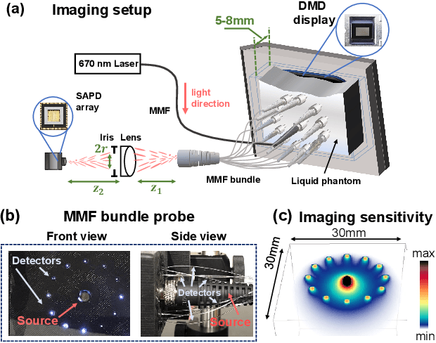
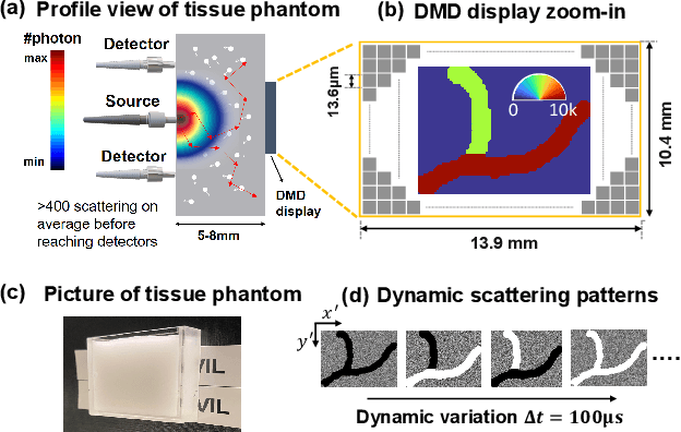
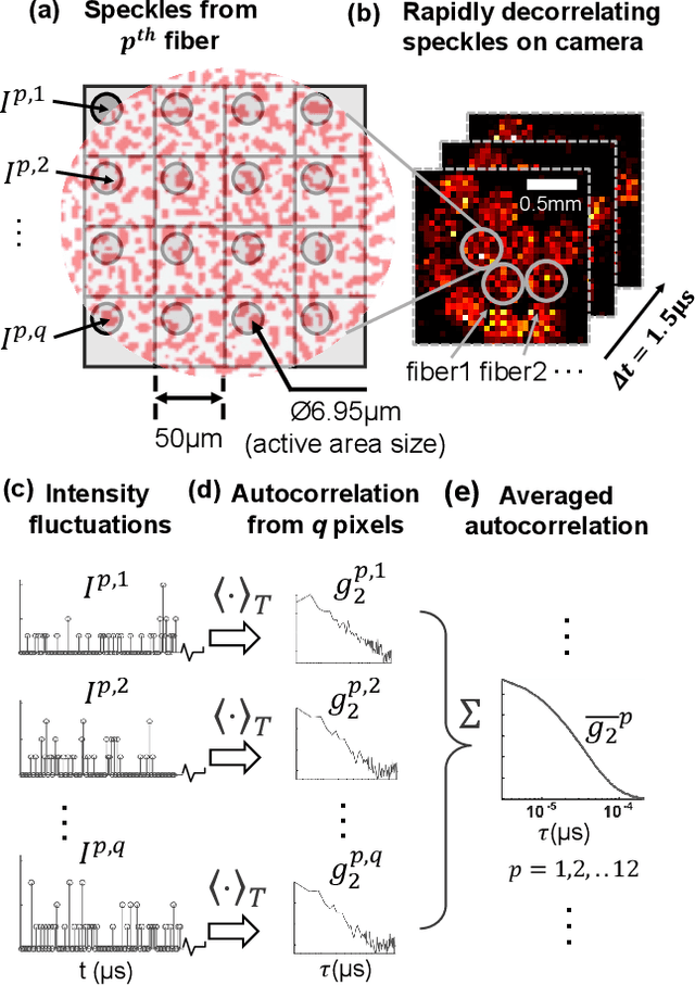
Noninvasive optical imaging through dynamic scattering media has numerous important biomedical applications but still remains a challenging task. While standard methods aim to form images based upon optical absorption or fluorescent emission, it is also well-established that the temporal correlation of scattered coherent light diffuses through tissue much like optical intensity. Few works to date, however, have aimed to experimentally measure and process such data to demonstrate deep-tissue imaging of decorrelation dynamics. In this work, we take advantage of a single-photon avalanche diode (SPAD) array camera, with over one thousand detectors, to simultaneously detect speckle fluctuations at the single-photon level from 12 different phantom tissue surface locations delivered via a customized fiber bundle array. We then apply a deep neural network to convert the acquired single-photon measurements into video of scattering dynamics beneath rapidly decorrelating liquid tissue phantoms. We demonstrate the ability to record video of dynamic events occurring 5-8 mm beneath a decorrelating tissue phantom with mm-scale resolution and at a 2.5-10 Hz frame rate.
Mesoscopic photogrammetry with an unstabilized phone camera
Dec 11, 2020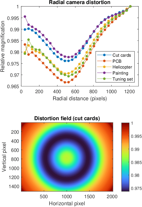
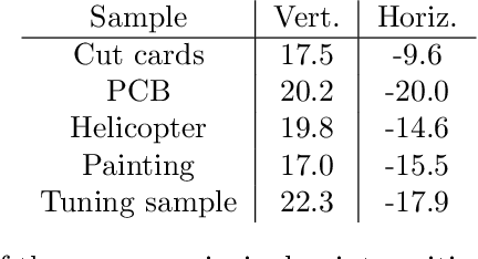
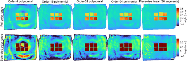
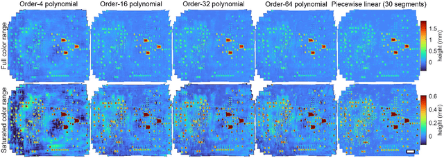
We present a feature-free photogrammetric technique that enables quantitative 3D mesoscopic (mm-scale height variation) imaging with tens-of-micron accuracy from sequences of images acquired by a smartphone at close range (several cm) under freehand motion without additional hardware. Our end-to-end, pixel-intensity-based approach jointly registers and stitches all the images by estimating a coaligned height map, which acts as a pixel-wise radial deformation field that orthorectifies each camera image to allow homographic registration. The height maps themselves are reparameterized as the output of an untrained encoder-decoder convolutional neural network (CNN) with the raw camera images as the input, which effectively removes many reconstruction artifacts. Our method also jointly estimates both the camera's dynamic 6D pose and its distortion using a nonparametric model, the latter of which is especially important in mesoscopic applications when using cameras not designed for imaging at short working distances, such as smartphone cameras. We also propose strategies for reducing computation time and memory, applicable to other multi-frame registration problems. Finally, we demonstrate our method using sequences of multi-megapixel images captured by an unstabilized smartphone on a variety of samples (e.g., painting brushstrokes, circuit board, seeds).
Physics-enhanced machine learning for virtual fluorescence microscopy
Apr 21, 2020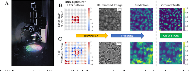
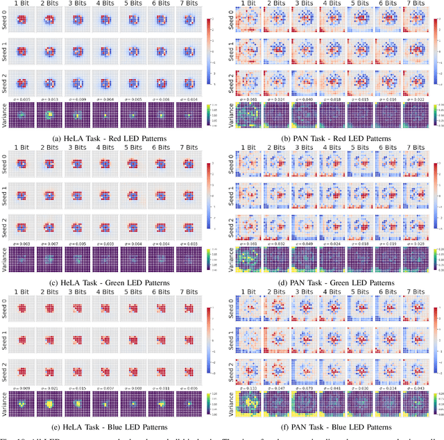
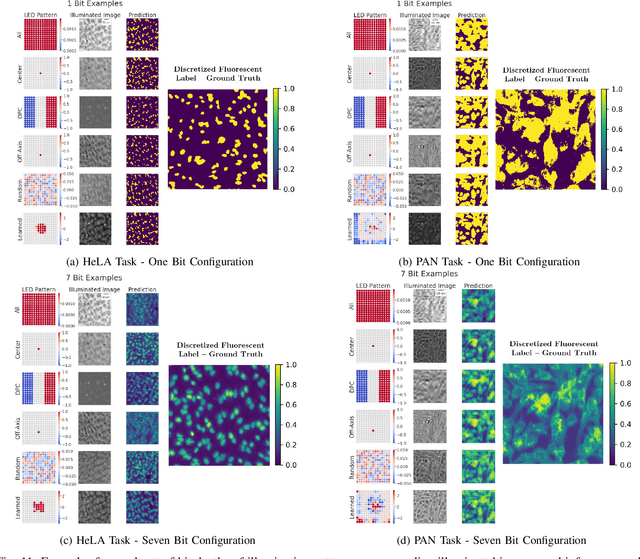
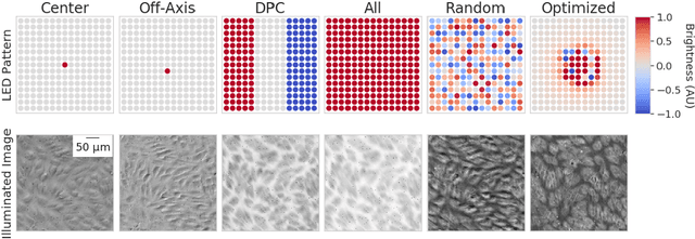
This paper introduces a new method of data-driven microscope design for virtual fluorescence microscopy. Our results show that by including a model of illumination within the first layers of a deep convolutional neural network, it is possible to learn task-specific LED patterns that substantially improve the ability to infer fluorescence image information from unstained transmission microscopy images. We validated our method on two different experimental setups, with different magnifications and different sample types, to show a consistent improvement in performance as compared to conventional illumination methods. Additionally, to understand the importance of learned illumination on inference task, we varied the dynamic range of the fluorescent image targets (from one to seven bits), and showed that the margin of improvement for learned patterns increased with the information content of the target. This work demonstrates the power of programmable optical elements at enabling better machine learning algorithm performance and at providing physical insight into next generation of machine-controlled imaging systems.