 Add to Chrome
 Add to Firefox
 Add to Edge
Unsupervised detection of small hyperreflective features in ultrahigh resolution optical coherence tomography
Mar 26, 2023Recent advances in optical coherence tomography such as the development of high speed ultrahigh resolution scanners and corresponding signal processing techniques may reveal new potential biomarkers in retinal diseases. Newly visible features are, for example, small hyperreflective specks in age-related macular degeneration. Identifying these new markers is crucial to investigate potential association with disease progression and treatment outcomes. Therefore, it is necessary to reliably detect these features in 3D volumetric scans. Because manual labeling of entire volumes is infeasible a need for automatic detection arises. Labeled datasets are often not publicly available and there are usually large variations in scan protocols and scanner types. Thus, this work focuses on an unsupervised approach that is based on local peak-detection and random walker segmentation to detect small features on each B-scan of the volume.
Retinal blood flow speed quantification at the capillary level using temporal autocorrelation fitting OCTA
Feb 22, 2023


Optical coherence tomography angiography (OCTA) can visualize vasculature structures, but provides limited information about the blood flow speeds. Here, we present a second generation variable interscan time analysis (VISTA) OCTA, which evaluates a quantitative surrogate marker for blood flow speed in vasculature. At the capillary level, spatially compiled OCTA and a simple temporal autocorrelation model, {\rho}({\tau}) = exp(-{\alpha}{\tau}), were used to evaluate a temporal autocorrelation decay constant, {\alpha}, as the blood flow speed marker. A 600 kHz A-scan rate swept-source provides short interscan time OCTA and fine A-scan spacing acquisition, while maintaining multi mm2 field of views for human retinal imaging. We demonstrate the cardiac pulsatility and repeatability of {\alpha} measured with VISTA. We show different {\alpha} for different retinal capillary plexuses in healthy eyes and present representative VISTA OCTA of eyes with diabetic retinopathy.
Maximum a posteriori signal recovery for optical coherence tomography angiography image generation and denoising
Oct 29, 2020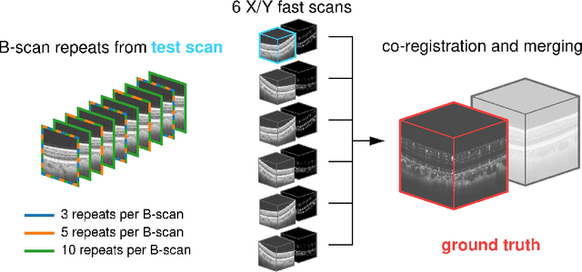
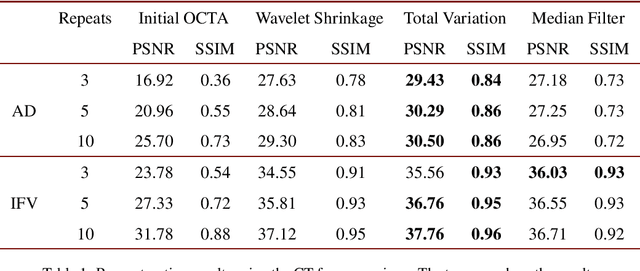
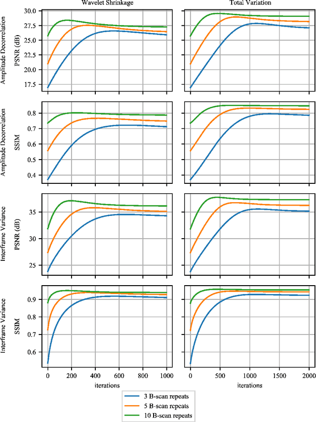
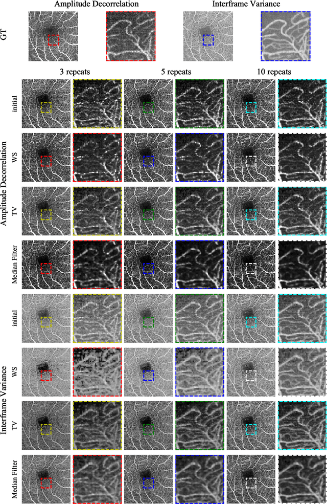
Optical coherence tomography angiography (OCTA) is a novel and clinically promising imaging modality to image retinal and sub-retinal vasculature. Based on repeated optical coherence tomography (OCT) scans, intensity changes are observed over time and used to compute OCTA image data. OCTA data are prone to noise and artifacts caused by variations in flow speed and patient movement. We propose a novel iterative maximum a posteriori signal recovery algorithm in order to generate OCTA volumes with reduced noise and increased image quality. This algorithm is based on previous work on probabilistic OCTA signal models and maximum likelihood estimates. Reconstruction results using total variation minimization and wavelet shrinkage for regularization were compared against an OCTA ground truth volume, merged from six co-registered single OCTA volumes. The results show a significant improvement in peak signal-to-noise ratio and structural similarity. The presented algorithm brings together OCTA image generation and Bayesian statistics and can be developed into new OCTA image generation and denoising algorithms.
Efficient and high accuracy 3-D OCT angiography motion correction in pathology
Oct 14, 2020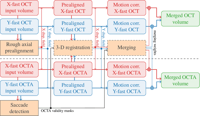
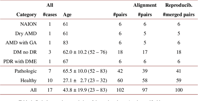
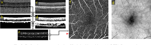
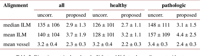
We propose a novel method for non-rigid 3-D motion correction of orthogonally raster-scanned optical coherence tomography angiography volumes. This is the first approach that aligns predominantly axial structural features like retinal layers and transverse angiographic vascular features in a joint optimization. Combined with the use of orthogonal scans and favorization of kinematically more plausible displacements, the approach allows subpixel alignment and micrometer-scale distortion correction in all 3 dimensions. As no specific structures or layers are segmented, the approach is by design robust to pathologic changes. It is furthermore designed for highly parallel implementation and brief runtime, allowing its integration in clinical routine even for high density or wide-field scans. We evaluated the algorithm with metrics related to clinically relevant features in a large-scale quantitative evaluation based on 204 volumetric scans of 17 subjects including both a wide range of pathologies and healthy controls. Using this method, we achieve state-of-the-art axial performance and show significant advances in both transverse co-alignment and distortion correction, especially in the pathologic subgroup.
Deep OCT Angiography Image Generation for Motion Artifact Suppression
Jan 08, 2020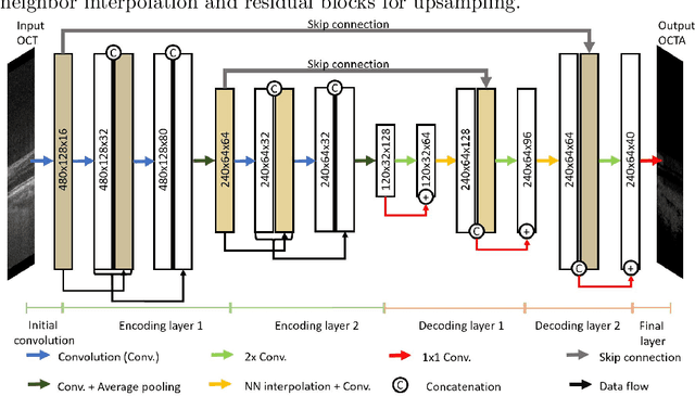
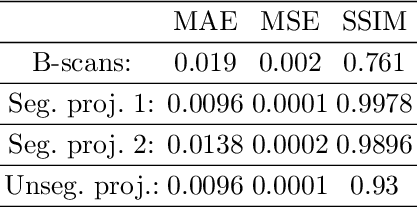
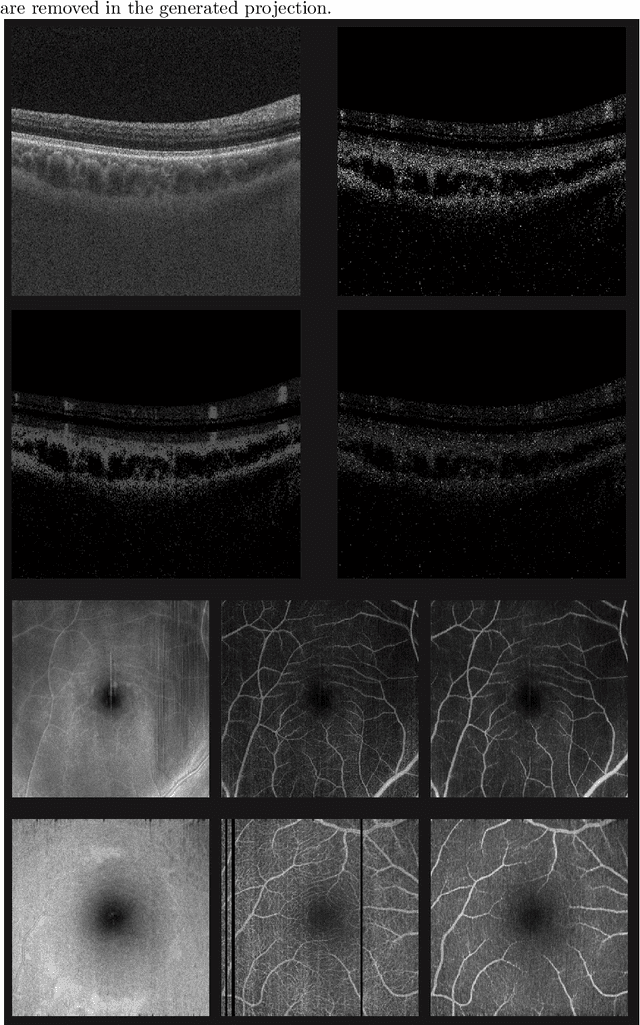
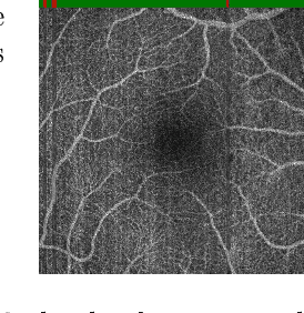
Eye movements, blinking and other motion during the acquisition of optical coherence tomography (OCT) can lead to artifacts, when processed to OCT angiography (OCTA) images. Affected scans emerge as high intensity (white) or missing (black) regions, resulting in lost information. The aim of this research is to fill these gaps using a deep generative model for OCT to OCTA image translation relying on a single intact OCT scan. Therefore, a U-Net is trained to extract the angiographic information from OCT patches. At inference, a detection algorithm finds outlier OCTA scans based on their surroundings, which are then replaced by the trained network. We show that generative models can augment the missing scans. The augmented volumes could then be used for 3-D segmentation or increase the diagnostic value.
Temporal and Volumetric Denoising via Quantile Sparse Image (QuaSI) Prior in Optical Coherence Tomography and Beyond
Jul 05, 2018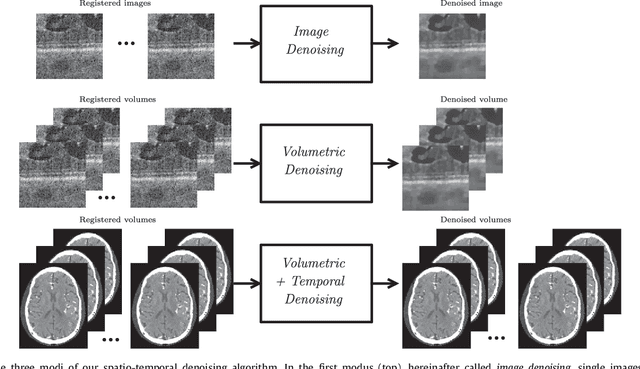
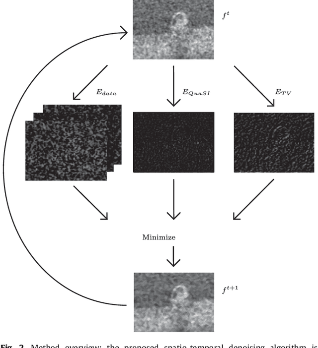
This paper introduces an universal and structure-preserving regularization term, called quantile sparse image (QuaSI) prior. The prior is suitable for denoising images from various medical imaging modalities. We demonstrate its effectiveness on volumetric optical coherence tomography (OCT) and computed tomography (CT) data, which show different noise and image characteristics. OCT offers high-resolution scans of the human retina but is inherently impaired by speckle noise. CT on the other hand has a lower resolution and shows high-frequency noise. For the purpose of denoising, we propose a variational framework based on the QuaSI prior and a Huber data fidelity model that can handle 3-D and 3-D+t data. Efficient optimization is facilitated through the use of an alternating direction method of multipliers (ADMM) scheme and the linearization of the quantile filter. Experiments on multiple datasets emphasize the excellent performance of the proposed method.
QuaSI: Quantile Sparse Image Prior for Spatio-Temporal Denoising of Retinal OCT Data
Mar 08, 2017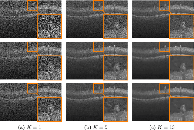
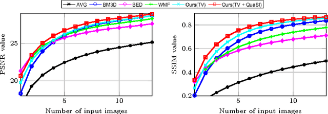
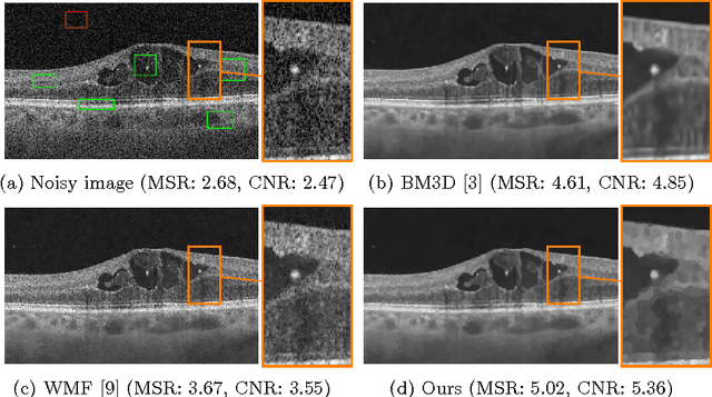
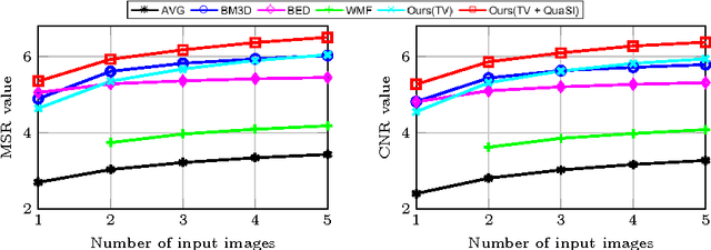
Optical coherence tomography (OCT) enables high-resolution and non-invasive 3D imaging of the human retina but is inherently impaired by speckle noise. This paper introduces a spatio-temporal denoising algorithm for OCT data on a B-scan level using a novel quantile sparse image (QuaSI) prior. To remove speckle noise while preserving image structures of diagnostic relevance, we implement our QuaSI prior via median filter regularization coupled with a Huber data fidelity model in a variational approach. For efficient energy minimization, we develop an alternating direction method of multipliers (ADMM) scheme using a linearization of median filtering. Our spatio-temporal method can handle both, denoising of single B-scans and temporally consecutive B-scans, to gain volumetric OCT data with enhanced signal-to-noise ratio. Our algorithm based on 4 B-scans only achieved comparable performance to averaging 13 B-scans and outperformed other current denoising methods.