 Add to Chrome
 Add to Firefox
 Add to Edge
Task-Adaptive 3D Cross-Field MRI Translation via Field-Conditioned Content-Style Pretraining
Aug 10, 2026
Magnetic field strength is a major source of domain shift in magnetic resonance imaging (MRI), affecting signal-to-noise ratio, tissue contrast, spatial detail, and the visibility of anatomical boundaries. The MRIxFields 2026 challenge investigates this problem through cross-field MRI translation across acquisitions at 0.1T, 1.5T, 3T, 5T, and 7T. Its three tasks, Any-to-7T, 0.1T-to-High, and Any-to-Any synthesis, require the generation of target-field image characteristics while preserving subject-specific anatomy. This problem is particularly challenging because paired acquisitions of the same subject across multiple field strengths are rarely available for training. We propose a 3D unpaired cross-field MRI translation framework based on field-conditioned content-style pretraining. The proposed framework first learns controllable field-to-field translation across all available field strengths by disentangling anatomical content from field-dependent contrast characteristics. The pretrained backbone is then adapted to task-specific target domains. Our model comprises a 3D content encoder, a 3D style encoder, a field-conditioned style generator, an AdaIN-modulated decoder, and a multi-field discriminator. Adversarial learning encourages realistic target-field appearance, while cycle-consistency, identity, content, style, and diversity constraints promote anatomical fidelity and controllable translation. We evaluate the proposed method on MRIxFields data spanning five field strengths and three MRI modalities. Experiments on paired test data demonstrate that the framework can adapt to the three challenge settings while preserving three-dimensional anatomical structure in the synthesized volumes. The implementation code is publicly available at https://github.com/Idea89560041/3D-MRI-Field-Translation.

MedARC: Training-Free Adaptive Redundancy Compression of Visual Tokens for 3D Medical Vision-Language Models
Jul 29, 2026Integrating 3D medical images with vision-language models (VLMs) holds substantial promise for computer-aided diagnosis. However, volumetric images generate prohibitively long visual-token sequences with considerable spatial and inter-slice redundancy. Existing token compression methods typically apply uniform reduction or rely on a single importance signal, increasing the risk of removing regions that are clinically relevant to the query or structurally distinctive. To address this limitation, we propose MedARC, a unified, training-free framework for Adaptive Redundancy Compression of visual tokens in 3D medical VLMs. MedARC estimates token importance by integrating three complementary cues: self-attention from the VLM vision encoder, which reflects the model's intrinsic visual focus; similarity between projected visual tokens and text embeddings, which identifies query-relevant regions; and deviations of local visual foundation model features from the volume-level feature center, which highlight structurally distinctive anatomy. The resulting importance distribution guides a saliency-aware merging strategy that preserves informative tokens while consolidating redundant ones rather than simply discarding them. Experiments on CT-RATE and MR-RATE show that MedARC reduces visual-token overhead and inference time while preserving or improving diagnostic performance. Its multi-cue scoring cost is outweighed by the savings from processing fewer tokens, with greater benefits expected for larger language models.
IHF-Harmony: Multi-Modality Magnetic Resonance Images Harmonization using Invertible Hierarchy Flow Model
Feb 25, 2026Retrospective MRI harmonization is limited by poor scalability across modalities and reliance on traveling subject datasets. To address these challenges, we introduce IHF-Harmony, a unified invertible hierarchy flow framework for multi-modality harmonization using unpaired data. By decomposing the translation process into reversible feature transformations, IHF-Harmony guarantees bijective mapping and lossless reconstruction to prevent anatomical distortion. Specifically, an invertible hierarchy flow (IHF) performs hierarchical subtractive coupling to progressively remove artefact-related features, while an artefact-aware normalization (AAN) employs anatomy-fixed feature modulation to accurately transfer target characteristics. Combined with anatomy and artefact consistency loss objectives, IHF-Harmony achieves high-fidelity harmonization that retains source anatomy. Experiments across multiple MRI modalities demonstrate that IHF-Harmony outperforms existing methods in both anatomical fidelity and downstream task performance, facilitating robust harmonization for large-scale multi-site imaging studies. Code will be released upon acceptance.
Advancing Brain Tumor Inpainting with Generative Models
Feb 02, 2024Synthesizing healthy brain scans from diseased brain scans offers a potential solution to address the limitations of general-purpose algorithms, such as tissue segmentation and brain extraction algorithms, which may not effectively handle diseased images. We consider this a 3D inpainting task and investigate the adaptation of 2D inpainting methods to meet the requirements of 3D magnetic resonance imaging(MRI) data. Our contributions encompass potential modifications tailored to MRI-specific needs, and we conducted evaluations of multiple inpainting techniques using the BraTS2023 Inpainting datasets to assess their efficacy and limitations.
Two-stage Contextual Transformer-based Convolutional Neural Network for Airway Extraction from CT Images
Dec 15, 2022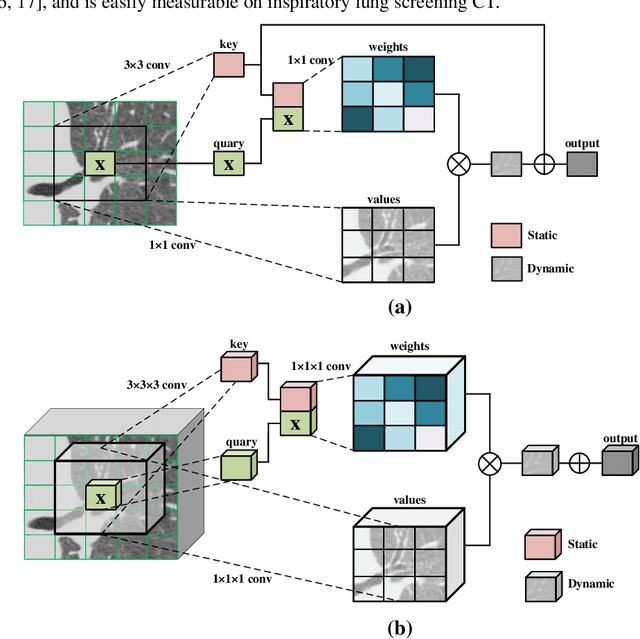

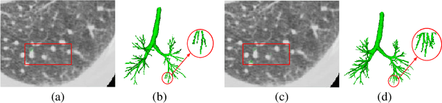

Accurate airway extraction from computed tomography (CT) images is a critical step for planning navigation bronchoscopy and quantitative assessment of airway-related chronic obstructive pulmonary disease (COPD). The existing methods are challenging to sufficiently segment the airway, especially the high-generation airway, with the constraint of the limited label and cannot meet the clinical use in COPD. We propose a novel two-stage 3D contextual transformer-based U-Net for airway segmentation using CT images. The method consists of two stages, performing initial and refined airway segmentation. The two-stage model shares the same subnetwork with different airway masks as input. Contextual transformer block is performed both in the encoder and decoder path of the subnetwork to finish high-quality airway segmentation effectively. In the first stage, the total airway mask and CT images are provided to the subnetwork, and the intrapulmonary airway mask and corresponding CT scans to the subnetwork in the second stage. Then the predictions of the two-stage method are merged as the final prediction. Extensive experiments were performed on in-house and multiple public datasets. Quantitative and qualitative analysis demonstrate that our proposed method extracted much more branches and lengths of the tree while accomplishing state-of-the-art airway segmentation performance. The code is available at https://github.com/zhaozsq/airway_segmentation.