 Add to Chrome
 Add to Firefox
 Add to Edge
The Next Layer: Augmenting Foundation Models with Structure-Preserving and Attention-Guided Learning for Local Patches to Global Context Awareness in Computational Pathology
Aug 27, 2025


Foundation models have recently emerged as powerful feature extractors in computational pathology, yet they typically omit mechanisms for leveraging the global spatial structure of tissues and the local contextual relationships among diagnostically relevant regions - key elements for understanding the tumor microenvironment. Multiple instance learning (MIL) remains an essential next step following foundation model, designing a framework to aggregate patch-level features into slide-level predictions. We present EAGLE-Net, a structure-preserving, attention-guided MIL architecture designed to augment prediction and interpretability. EAGLE-Net integrates multi-scale absolute spatial encoding to capture global tissue architecture, a top-K neighborhood-aware loss to focus attention on local microenvironments, and background suppression loss to minimize false positives. We benchmarked EAGLE-Net on large pan-cancer datasets, including three cancer types for classification (10,260 slides) and seven cancer types for survival prediction (4,172 slides), using three distinct histology foundation backbones (REMEDIES, Uni-V1, Uni2-h). Across tasks, EAGLE-Net achieved up to 3% higher classification accuracy and the top concordance indices in 6 of 7 cancer types, producing smooth, biologically coherent attention maps that aligned with expert annotations and highlighted invasive fronts, necrosis, and immune infiltration. These results position EAGLE-Net as a generalizable, interpretable framework that complements foundation models, enabling improved biomarker discovery, prognostic modeling, and clinical decision support
Multi-scale Regional Attention Deeplab3+: Multiple Myeloma Plasma Cells Segmentation in Microscopic Images
May 13, 2021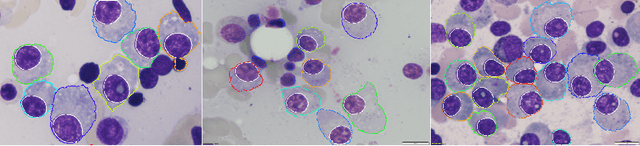
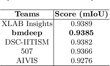
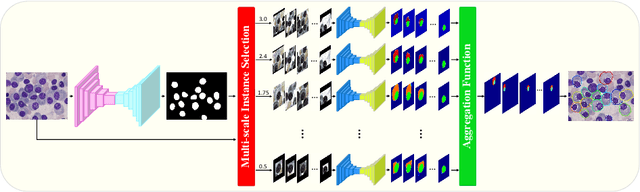
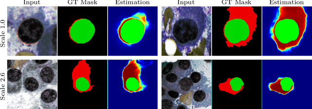
Multiple myeloma cancer is a type of blood cancer that happens when the growth of abnormal plasma cells becomes out of control in the bone marrow. There are various ways to diagnose multiple myeloma in bone marrow such as complete blood count test (CBC) or counting myeloma plasma cell in aspirate slide images using manual visualization or through image processing technique. In this work, an automatic deep learning method for the detection and segmentation of multiple myeloma plasma cell have been explored. To this end, a two-stage deep learning method is designed. In the first stage, the nucleus detection network is utilized to extract each instance of a cell of interest. The extracted instance is then fed to the multi-scale function to generate a multi-scale representation. The objective of the multi-scale function is to capture the shape variation and reduce the effect of object scale on the cytoplasm segmentation network. The generated scales are then fed into a pyramid of cytoplasm networks to learn the segmentation map in various scales. On top of the cytoplasm segmentation network, we included a scale aggregation function to refine and generate a final prediction. The proposed approach has been evaluated on the SegPC2021 grand-challenge and ranked second on the final test phase among all teams.