 Add to Chrome
 Add to Firefox
 Add to Edge
ProSAC-CT: Progressive Spectral-Anatomical Co-Guided Multi-Stage Diffusion Model for Low-Dose CT Denoising
Jul 02, 2026
Low-dose computed tomography (LDCT) reduces radiation exposure but introduces stronger quantum noise, streak artifacts, and local texture degradation, which can obscure anatomical boundaries and weaken low-contrast structures. Diffusion models are promising for LDCT denoising by progressively recovering normal-dose CT (NDCT) images from degraded LDCT inputs, but existing methods often suffer from insufficient anatomical guidance, uncertain frequency-dependent recovery, and uniform reverse-process modeling. We propose ProSAC-CT, a progressive spectral-anatomical co-guided multi-stage diffusion model for image-domain LDCT denoising. ProSAC-CT integrates an anatomical-prior-guided conditioning (APGC) module, a residual frequency-domain decoupling stage (RFDDS), and a time-step-decoupling denoising decoder (TD3). APGC extracts LDCT-derived structural guidance, RFDDS enhances frequency-aware representations, and TD3 assigns them to different reverse-diffusion stages for anatomical stabilization, boundary refinement, and fine-detail recovery. Experiments on four LDCT degradation benchmarks show that ProSAC-CT improves image fidelity, structural similarity, perceptual quality, and information preservation over representative methods while better preserving boundary-sensitive anatomical details. Downstream anatomical-region classification on Mayo-2020 further indicates that ProSAC-CT retains task-relevant anatomical information, supporting its practical use for low-dose CT denoising.

In-context learning for medical image segmentation
Dec 17, 2024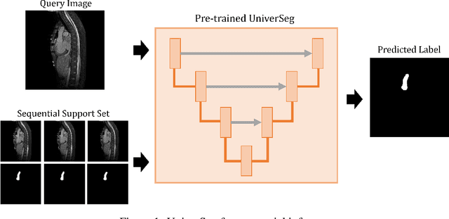
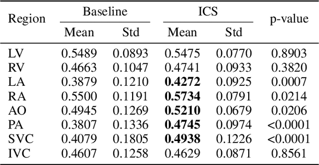

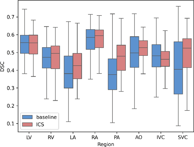
Annotation of medical images, such as MRI and CT scans, is crucial for evaluating treatment efficacy and planning radiotherapy. However, the extensive workload of medical professionals limits their ability to annotate large image datasets, posing a bottleneck for AI applications in medical imaging. To address this, we propose In-context Cascade Segmentation (ICS), a novel method that minimizes annotation requirements while achieving high segmentation accuracy for sequential medical images. ICS builds on the UniverSeg framework, which performs few-shot segmentation using support images without additional training. By iteratively adding the inference results of each slice to the support set, ICS propagates information forward and backward through the sequence, ensuring inter-slice consistency. We evaluate the proposed method on the HVSMR dataset, which includes segmentation tasks for eight cardiac regions. Experimental results demonstrate that ICS significantly improves segmentation performance in complex anatomical regions, particularly in maintaining boundary consistency across slices, compared to baseline methods. The study also highlights the impact of the number and position of initial support slices on segmentation accuracy. ICS offers a promising solution for reducing annotation burdens while delivering robust segmentation results, paving the way for its broader adoption in clinical and research applications.
PlaNet-S: Automatic Semantic Segmentation of Placenta
Dec 18, 2023[Purpose] To develop a fully automated semantic placenta segmentation model that integrates the U-Net and SegNeXt architectures through ensemble learning. [Methods] A total of 218 pregnant women with suspected placental anomalies who underwent magnetic resonance imaging (MRI) were enrolled, yielding 1090 annotated images for developing a deep learning model for placental segmentation. The images were standardized and divided into training and test sets. The performance of PlaNet-S, which integrates U-Net and SegNeXt within an ensemble framework, was assessed using Intersection over Union (IoU) and counting connected components (CCC) against the U-Net model. [Results] PlaNet-S had significantly higher IoU (0.73 +/- 0.13) than that of U-Net (0.78 +/- 0.010) (p<0.01). The CCC for PlaNet-S was significantly higher than that for U-Net (p<0.01), matching the ground truth in 86.0\% and 56.7\% of the cases, respectively. [Conclusion]PlaNet-S performed better than the traditional U-Net in placental segmentation tasks. This model addresses the challenges of time-consuming physician-assisted manual segmentation and offers the potential for diverse applications in placental imaging analyses.