 Add to Chrome
 Add to Firefox
 Add to Edge
Trust but Verify: Mitigating Medical Hallucinations via Post-Hoc Adversarial Auditing and Multi-Agent Feedback Loops
Jun 12, 2026
Large Language Models (LLMs) are increasingly deployed in healthcare settings, yet their tendency to hallucinate poses risks when clinical decisions are involved. This study examine whether LLMs recommend recently banned or withdrawn pharmaceuticals when answering clinical questions and tests an agent-based method for reducing such errors. We developed a five-agent "Trust but Verify" system using a single LLM backbone. To measure regulatory knowledge obsolescence, we created an adversarial dataset of 103 clinical MCQs where historically correct answers now refer to banned substances. This scale ensures statistical significance across various therapeutic classes. We evaluated three open-access model families (GPT-OSS, Llama-3, Falcon-3) under vanilla and agentic conditions. Performance was measured via pointwise score, label accuracy, Hallucination Error Rate (HER), and Component Fidelity (CF) score. We also observed clinical safety regression in proprietary models. In default configurations, all models showed high hallucination rates, consistently selecting banned drugs that matched training data patterns. Our proposed agentic architecture reduced HER by approximately 53% across models. Pointwise scores shifted from -0.25 (unsafe recommendation) toward 0.0 (appropriate refusal). The safety audit intercepted dangerous outputs even when models' parametric knowledge favored the banned substance. The proposed multi-agent framework offers a model-agnostic method for enforcing regulatory compliance that prioritizes patient safety over fluent text generation. Our work demonstrates a practical approach for deploying autonomous AI systems in safety-critical healthcare settings. It shows how real-time regulatory data can be integrated into LLM pipelines to support clinical decision-making.

Pixel Wised Lesion Prediction on COVID-19 CT Imagery: A Comparative Analysis of Automated Image Segmentation Architectures
May 19, 2026In recent years, there has been a notable increase in the level of attention that is given to algorithms based on deep learning in the context of medical image segmentation. Nevertheless, the reliability of the field has been hindered due to the absence of a standardized methodology for performance analysis and the utilization of different datasets in previous research. The primary objective of the research is to comprehensively evaluate contemporary segmentation frameworks combined with state-of-the-art pre-trained backbones in order to accurately predict COVID-19 lesions in CT images. Moreover, this evaluation can serve as a point of reference for the segmentation of images in various other imaging scenarios. In order to accomplish this, we integrate four distinct deep learning architectures, namely Unet, PSPNet, Linknet, and FPN, with six pre-trained encoders, including VGG 19, DenseNet 121, Inception ResNet V2, MobileNet V2, SeresNet 101, and EfficientNet B0. This approach enables the development of diverse testing architectures. In the context of image segmentation, our research encompassed both binary and multi-class experimentation. The findings derived from our analysis of three distinct COVID-19 CT segmentation datasets indicate that deep learning architectures yield precise and efficient segmentation outcomes. Significantly, a maximum F1-Score of 98% was attained for binary class segmentation, while multi-class segmentation yielded F1-Scores of 75% and 77% across two separate datasets. The utilization of artificial intelligence and deep learning enhances the diagnostic process for pandemic diseases across multiple dimensions.
A Comprehensive Comparison of Deep Learning Architectures for COVID-19 Classification on CT & X-ray Imagery
May 19, 2026COVID-19 was a significant challenge that led to the loss of numerous lives daily. Not only a certain country was involved in this outbreak, but even the world has suffered because of the coronavirus. Imaging techniques using computed tomography (CT) and X-rays of the lungs are the most useful tools for the COVID-19 or any other pandemic disease screening process. Technology today has revolutionized the world by using artificial intelligence to replace manual processes with automated machines, which enable the system to imitate the human brain by making wise decisions based on experience. Motivated by this, our work proposes to use convolutional neural networks (CNN) based models for designing a computer-aided diagnosis (CAD) system that differentiates between COVID-19 and healthy lung pictures. We used two different sets of X-ray images of the lungs in addition to two different sets of CT scans and the classification is done using a variety of networks that have been pre-trained such as VGG (16, 19), Densenet (121), Resnet (50, 50 V2, 101 V2), Mobile net (V2), Xception Inception (V3, Resnet V2), Efficient net (B0) and Nasnet (Large). On the X-ray and CT image datasets, Resnet and VGG architecture have shown the ability to properly differentiate COVID-19 from normal images, with an average accuracy of 95 to 98 percent respectively. Our acquired results on the classification datasets are competitive and superior to previously reported findings in the literature.
Classification of White Blood Cells Using Machine and Deep Learning Models: A Systematic Review
Aug 21, 2023Machine learning (ML) and deep learning (DL) models have been employed to significantly improve analyses of medical imagery, with these approaches used to enhance the accuracy of prediction and classification. Model predictions and classifications assist diagnoses of various cancers and tumors. This review presents an in-depth analysis of modern techniques applied within the domain of medical image analysis for white blood cell classification. The methodologies that use blood smear images, magnetic resonance imaging (MRI), X-rays, and similar medical imaging domains are identified and discussed, with a detailed analysis of ML/DL techniques applied to the classification of white blood cells (WBCs) representing the primary focus of the review. The data utilized in this research has been extracted from a collection of 136 primary papers that were published between the years 2006 and 2023. The most widely used techniques and best-performing white blood cell classification methods are identified. While the use of ML and DL for white blood cell classification has concurrently increased and improved in recent year, significant challenges remain - 1) Availability of appropriate datasets remain the primary challenge, and may be resolved using data augmentation techniques. 2) Medical training of researchers is recommended to improve current understanding of white blood cell structure and subsequent selection of appropriate classification models. 3) Advanced DL networks including Generative Adversarial Networks, R-CNN, Fast R-CNN, and faster R-CNN will likely be increasingly employed to supplement or replace current techniques.
A Hybrid VDV Model for Automatic Diagnosis of Pneumothorax using Class-Imbalanced Chest X-rays Dataset
Dec 22, 2020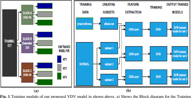
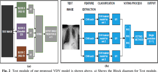
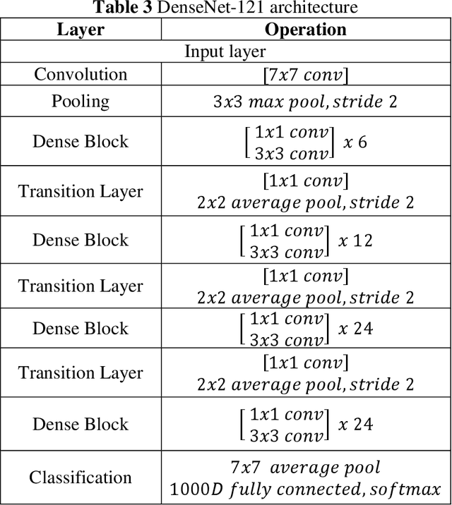
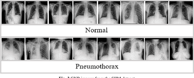
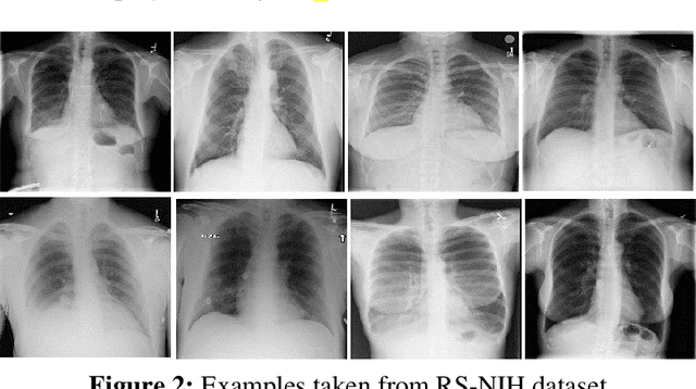
Pneumothorax, a life threatening disease, needs to be diagnosed immediately and efficiently. The prognosis in this case is not only time consuming but also prone to human errors. So an automatic way of accurate diagnosis using chest X-rays is the utmost requirement. To-date, most of the available medical images datasets have class-imbalance issue. The main theme of this study is to solve this problem along with proposing an automated way of detecting pneumothorax. We first compare the existing approaches to tackle the class-imbalance issue and find that data-level-ensemble (i.e. ensemble of subsets of dataset) outperforms other approaches. Thus, we propose a novel framework named as VDV model, which is a complex model-level-ensemble of data-level-ensembles and uses three convolutional neural networks (CNN) including VGG16, VGG-19 and DenseNet-121 as fixed feature extractors. In each data-level-ensemble features extracted from one of the pre-defined CNN are fed to support vector machine (SVM) classifier, and output from each data-level-ensemble is calculated using voting method. Once outputs from the three data-level-ensembles with three different CNN architectures are obtained, then, again, voting method is used to calculate the final prediction. Our proposed framework is tested on SIIM ACR Pneumothorax dataset and Random Sample of NIH Chest X-ray dataset (RS-NIH). For the first dataset, 85.17% Recall with 86.0% Area under the Receiver Operating Characteristic curve (AUC) is attained. For the second dataset, 90.9% Recall with 95.0% AUC is achieved with random split of data while 85.45% recall with 77.06% AUC is obtained with patient-wise split of data. For RS-NIH, the obtained results are higher as compared to previous results from literature However, for first dataset, direct comparison cannot be made, since this dataset has not been used earlier for Pneumothorax classification.
Automatic Diagnosis of Pneumothorax from Chest Radiographs: A Systematic Literature Review
Dec 21, 2020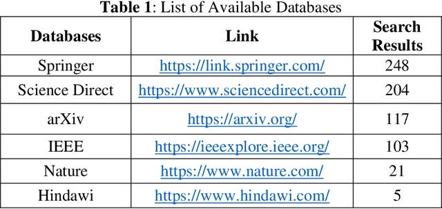
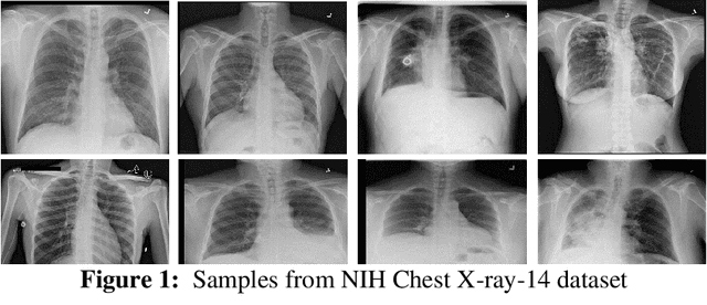
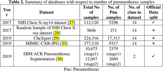
Among various medical imaging tools, chest radiographs are the most important and widely used diagnostic tool for detection of thoracic pathologies. Researches are being carried out in order to propose robust automatic diagnostic tool for detection of pathologies from chest radiographs. Artificial Intelligence techniques especially deep learning methodologies have been found to be giving promising results in automating the field of medicine. Lot of research has been done for automatic and fast detection of pneumothorax from chest radiographs while proposing several frameworks based on artificial intelligence and machine learning techniques. This study summarizes the existing literature for the automatic detection of pneumothorax from chest x-rays along with describing the available chest radiographs datasets. The comparative analysis of the literature is also provided in terms of goodness and limitations of the existing literature along with highlighting the research gaps which need to be further explored. The paper provides a brief overview of the present work for pneumothorax detection for helping the researchers in selection of optimal approach for future research.
Exploring Language-Independent Emotional Acoustic Features via Feature Selection
Sep 01, 2010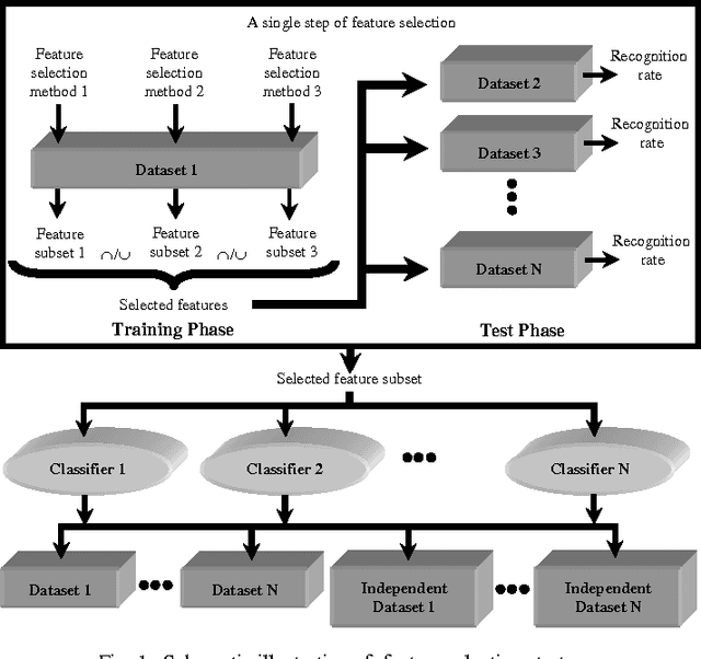
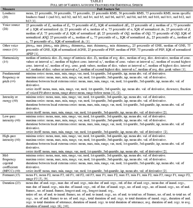
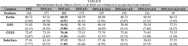
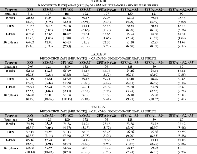
We propose a novel feature selection strategy to discover language-independent acoustic features that tend to be responsible for emotions regardless of languages, linguistics and other factors. Experimental results suggest that the language-independent feature subset discovered yields the performance comparable to the full feature set on various emotional speech corpora.
Emotional State Categorization from Speech: Machine vs. Human
Sep 01, 2010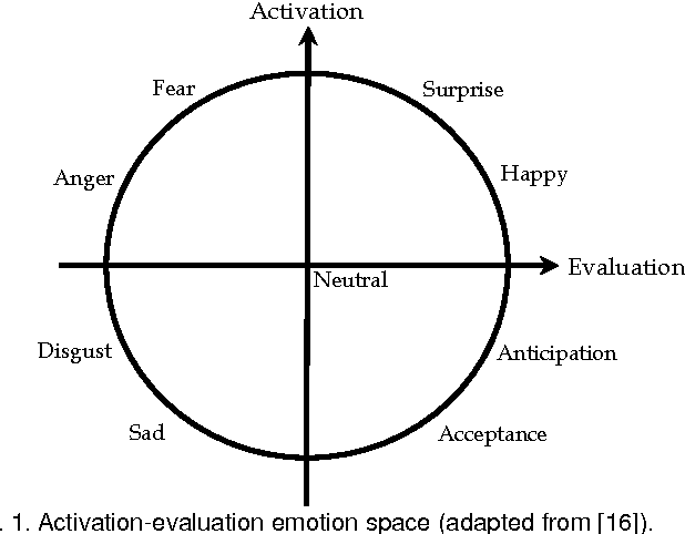
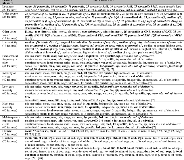
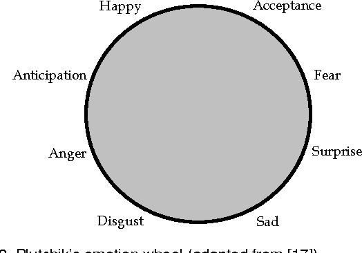
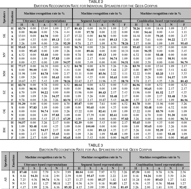
This paper presents our investigations on emotional state categorization from speech signals with a psychologically inspired computational model against human performance under the same experimental setup. Based on psychological studies, we propose a multistage categorization strategy which allows establishing an automatic categorization model flexibly for a given emotional speech categorization task. We apply the strategy to the Serbian Emotional Speech Corpus (GEES) and the Danish Emotional Speech Corpus (DES), where human performance was reported in previous psychological studies. Our work is the first attempt to apply machine learning to the GEES corpus where the human recognition rates were only available prior to our study. Unlike the previous work on the DES corpus, our work focuses on a comparison to human performance under the same experimental settings. Our studies suggest that psychology-inspired systems yield behaviours that, to a great extent, resemble what humans perceived and their performance is close to that of humans under the same experimental setup. Furthermore, our work also uncovers some differences between machine and humans in terms of emotional state recognition from speech.