 Add to Chrome
 Add to Firefox
 Add to Edge
MonoUNet: A Robust Tiny Neural Network for Automated Knee Cartilage Segmentation on Point-of-Care Ultrasound Devices
Apr 09, 2026
Objective: To develop a robust and compact deep learning model for automated knee cartilage segmentation on point-of-care ultrasound (POCUS) devices. Methods: We propose MonoUNet, an ultra-compact U-Net consisting of (i) an aggressively reduced backbone with an asymmetric decoder, (ii) a trainable monogenic block that extracts multi-scale local phase features, and (iii) a gated feature injection mechanism that integrates these features into the encoder stages to reduce sensitivity to variations in ultrasound image appearance and improve robustness across devices. MonoUNet was evaluated on a multi-site, multi-device knee cartilage ultrasound dataset acquired using cart-based, portable, and handheld POCUS devices. Results: Overall, MonoUNet outperformed existing lightweight segmentation models, with average Dice scores ranging from 92.62% to 94.82% and mean average surface distance (MASD) values between 0.133 mm and 0.254 mm. MonoUNet reduces the number of parameters by 10x--700x and computational cost by 14x--2000x relative to existing lightweight models. MonoUNet cartilage outcomes showed excellent reliability and agreement with the manual outcomes: intraclass correlation coefficients (ICC$_{2,k})$=0.96 and bias=2.00% (0.047 mm) for average thickness, and ICC$_{2,k}$=0.99 and bias=0.80% (0.328 a.u.) for echo intensity. Conclusion: Incorporating trainable local phase features improves the robustness of highly compact neural networks for knee cartilage segmentation across varying acquisition settings and could support scalable ultrasound-based assessment and monitoring of knee osteoarthritis using POCUS devices. The code is publicly available at https://github.com/alvinkimbowa/monounet.

Mono2D: A Trainable Monogenic Layer for Robust Knee Cartilage Segmentation on Out-of-Distribution 2D Ultrasound Data
Mar 12, 2025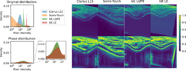
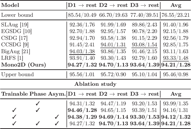
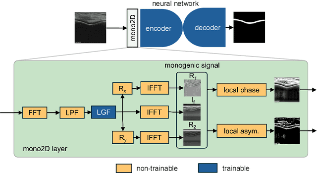
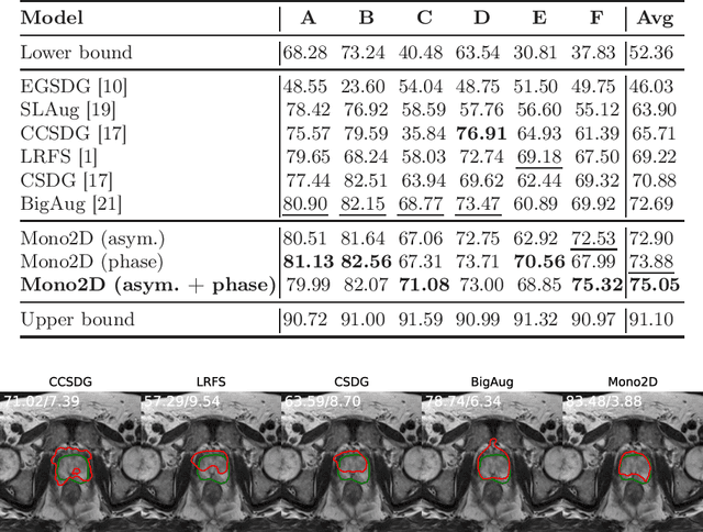
Automated knee cartilage segmentation using point-of-care ultrasound devices and deep-learning networks has the potential to enhance the management of knee osteoarthritis. However, segmentation algorithms often struggle with domain shifts caused by variations in ultrasound devices and acquisition parameters, limiting their generalizability. In this paper, we propose Mono2D, a monogenic layer that extracts multi-scale, contrast- and intensity-invariant local phase features using trainable bandpass quadrature filters. This layer mitigates domain shifts, improving generalization to out-of-distribution domains. Mono2D is integrated before the first layer of a segmentation network, and its parameters jointly trained alongside the network's parameters. We evaluated Mono2D on a multi-domain 2D ultrasound knee cartilage dataset for single-source domain generalization (SSDG). Our results demonstrate that Mono2D outperforms other SSDG methods in terms of Dice score and mean average surface distance. To further assess its generalizability, we evaluate Mono2D on a multi-site prostate MRI dataset, where it continues to outperform other SSDG methods, highlighting its potential to improve domain generalization in medical imaging. Nevertheless, further evaluation on diverse datasets is still necessary to assess its clinical utility.
Wireless vs. Traditional Ultrasound Assessed Knee Cartilage Outcomes Utilizing Automated Gain and Normalization Techniques
May 20, 2024Advancements in wireless ultrasound technology allow for point of care cartilage imaging, yet validation against traditional ultrasound units remains to be established for knee cartilage outcomes. Therefore, the purpose of our study was to establish the agreement of articular cartilage thickness and echo-intensity measures between traditional and wireless ultrasound units utilizing automatic-gain and normalization techniques. We used traditional and wireless ultrasound to assess the femoral cartilage via transverse suprapatellar scans with the knee in maximum flexion in 71 female NCAA Division I athletes (age: 20.0$\pm$1.3 years, height: 171.7$\pm$8.7 cm, mass: 69.4$\pm$11.0 kg). Wireless ultrasound images (auto-gain and standard gain) were compared to traditional ultrasound images (standard gain) before and after normalization. Ultrasound image pixel values were algebraically scaled to normalize imaging parameter differences between units. Mean thickness and echo-intensity of the global and sub-regions of interest were measured for unnormalized and normalized images. Intraclass correlation coefficients ($ICC_{2,k}$) for absolute agreement, standard error of the measurement, and minimum detectable difference were calculated between the traditional and wireless ultrasound units across both gain parameters and normalization. Cartilage thickness demonstrated good to excellent agreement for all regions ($ICC_{2,k} = 0.83 {\text -} 0.95$) regardless of gain and normalization. However, mean echo-intensity demonstrated poor to moderate agreement in all regions regardless of gain and normalization ($ICC_{2,k} = 0.23 {\text -} 0.68 $). While there was a high level of agreement between units when assessing cartilage thickness, further research in ultrasound beam forming may lead to improvements in agreement of cartilage echo-intensity between ultrasound units.