 Add to Chrome
 Add to Firefox
 Add to Edge
SAFViT: Spatial Attention Fusion Gating for Vision Transformer-Based Nucleus Segmentation and Classification
Jul 30, 2026
Accurate cell segmentation and classification are foundational to digital pathology, enabling quantitative tissue analysis for diagnosis and treatment planning. Encoder-decoder architectures that fuse multi-scale features through skip connections have become the dominant paradigm for this task, yet standard direct skip connections treat every spatial location equally, which leads to redundant and potentially conflicting information reaching the decoder. To overcome this problem, various gating mechanisms have been introduced, but most of them operate solely on filtering encoder information, neglecting the benefit of global contextual information from the decoder. This study proposes replacing conventional skip connections in a CellViT-based model with a novel Spatial Attention Fusion (SAF) Gating module. Each SAF gate concatenates the encoder skip and upsampled decoder features, compresses them through two pointwise convolutions with an intermediate ReLU, and applies a channel-wise softmax to produce a per-pixel "heatmap of trust" that sums to unity at every spatial location, allowing the network to learn where each source is most trustworthy. The resulting fused features improve the model's ability to detect the minority "Dead" class, which in turn enhances the multi-class panoptic quality (mPQ) on the PanNuke dataset. SAF Gating is compared against six gating alternatives including no gating, attention gates, squeeze-and-excitation, CBAM, cross-attention, and attentional feature fusion on PanNuke and MoNuSeg datasets. SAF Gating achieves the highest mPQ (0.471), a gain driven primarily by a 14.5-point improvement in Dead-class F1 score compared to ungated CellViT baseline.

Property-Constrained 3D Porous Media Reconstruction from 2D Images via Conditional Generative Adversarial Networks
Jul 02, 2026This study presents a conditional Generative Adversarial Network (cGAN) framework for generating 3D porous media volumes with controlled porosity, trained exclusively on 2D thin section images. The key innovation lies in combining property-conditioned generation with 2D-to-3D reconstruction, eliminating the need for expensive 3D training data while maintaining control over petrophysical properties. The framework employs a hybrid architecture with a 3D generator and 2D discriminator, where multi-axis slice extraction enables learning 3D-consistent structures from 2D training data. Porosity labels are extracted using an Enhanced U-Net segmentation model. The methodology was demonstrated on two carbonate samples with different lithologies: dolomite-anhydrite and pure dolomite. Results show that the framework successfully generates realistic 3D volumes capturing lithological features such as anhydrite inclusions and fine crystalline textures. Porosity control achieved an $R^2$ of 0.93, with mean absolute errors of 0.019 and 0.010 for the heterogeneous and homogeneous samples, respectively.
* 6 pages, 3 figures. Submitted to the 87th EAGE Annual Conference & Exhibition, Aberdeen, UK, June 2026
A Decade of Generative Adversarial Networks for Porous Material Reconstruction
Mar 12, 2026Digital reconstruction of porous materials has become increasingly critical for applications ranging from geological reservoir characterization to tissue engineering and electrochemical device design. While traditional methods such as micro-computed tomography and statistical reconstruction approaches have established foundations in this field, the emergence of deep learning techniques, particularly Generative Adversarial Networks (GANs), has revolutionized porous media reconstruction capabilities. This review systematically analyzes 96 peer-reviewed articles published from 2017 to early 2026, examining the evolution and applications of GAN-based approaches for porous material image reconstruction. We categorize GAN architectures into six distinct classes, namely Vanilla GANs, Multi-Scale GANs, Conditional GANs, Attention-Enhanced GANs, Style-based GANs, and Hybrid Architecture GANs. Our analysis reveals substantial progress including improvements in porosity accuracy (within 1% of original samples), permeability prediction (up to 79% reduction in mean relative errors), and achievable reconstruction volumes (from initial $64^3$ to current $2{,}200^3$ voxels). Despite these advances, persistent challenges remain in computational efficiency, memory constraints for large-scale reconstruction, and maintaining structural continuity in 2D-to-3D transformations. This systematic analysis provides a comprehensive framework for selecting appropriate GAN architectures based on specific application requirements.
DeepAngle: Fast calculation of contact angles in tomography images using deep learning
Nov 28, 2022DeepAngle is a machine learning-based method to determine the contact angles of different phases in the tomography images of porous materials. Measurement of angles in 3--D needs to be done within the surface perpendicular to the angle planes, and it could become inaccurate when dealing with the discretized space of the image voxels. A computationally intensive solution is to correlate and vectorize all surfaces using an adaptable grid, and then measure the angles within the desired planes. On the contrary, the present study provides a rapid and low-cost technique powered by deep learning to estimate the interfacial angles directly from images. DeepAngle is tested on both synthetic and realistic images against the direct measurement technique and found to improve the r-squared by 5 to 16% while lowering the computational cost 20 times. This rapid method is especially applicable for processing large tomography data and time-resolved images, which is computationally intensive. The developed code and the dataset are available at an open repository on GitHub (https://www.github.com/ArashRabbani/DeepAngle).
Automated segmentation and morphological characterization of placental histology images based on a single labeled image
Oct 07, 2022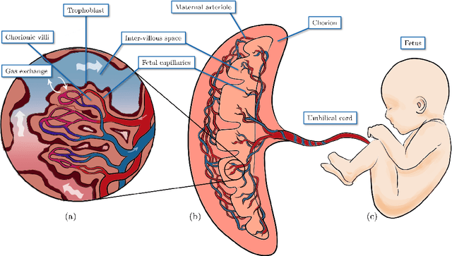
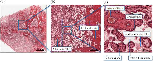
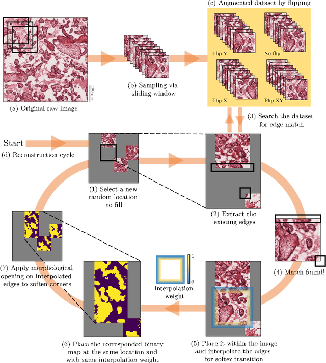
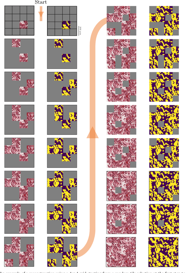
In this study, a novel method of data augmentation has been presented for the segmentation of placental histological images when the labeled data are scarce. This method generates new realizations of the placenta intervillous morphology while maintaining the general textures and orientations. As a result, a diversified artificial dataset of images is generated that can be used for training deep learning segmentation models. We have observed that on average the presented method of data augmentation led to a 42% decrease in the binary cross-entropy loss of the validation dataset compared to the common approach in the literature. Additionally, the morphology of the intervillous space is studied under the effect of the proposed image reconstruction technique, and the diversity of the artificially generated population is quantified. Due to the high resemblance of the generated images to the real ones, the applications of the proposed method may not be limited to placental histological images, and it is recommended that other types of tissues be investigated in future studies.
Temporal extrapolation of heart wall segmentation in cardiac magnetic resonance images via pixel tracking
Jul 30, 2022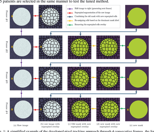
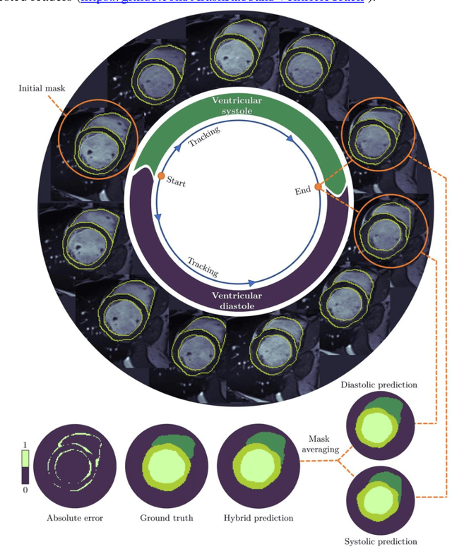
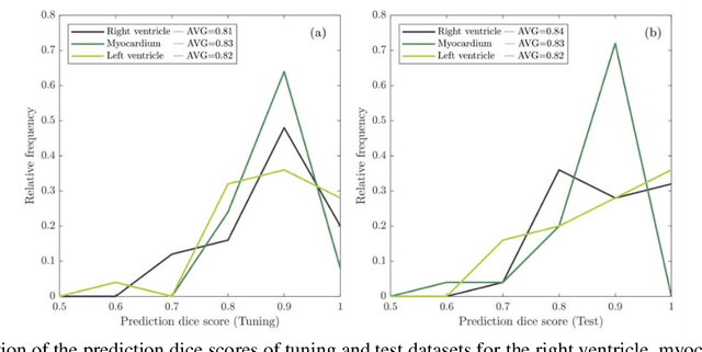
In this study, we have tailored a pixel tracking method for temporal extrapolation of the ventricular segmentation masks in cardiac magnetic resonance images. The pixel tracking process starts from the end-diastolic frame of the heart cycle using the available manually segmented images to predict the end-systolic segmentation mask. The superpixels approach is used to divide the raw images into smaller cells and in each time frame, new labels are assigned to the image cells which leads to tracking the movement of the heart wall elements through different frames. The tracked masks at the end of systole are compared with the already available manually segmented masks and dice scores are found to be between 0.81 to 0.84. Considering the fact that the proposed method does not necessarily require a training dataset, it could be an attractive alternative approach to deep learning segmentation methods in scenarios where training data are limited.
Resolution enhancement of placenta histological images using deep learning
Jul 30, 2022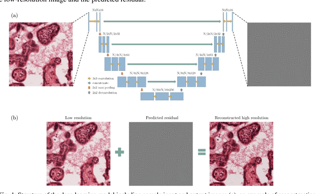
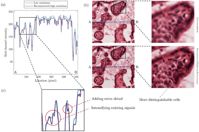
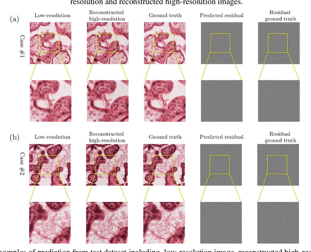
In this study, a method has been developed to improve the resolution of histological human placenta images. For this purpose, a paired series of high- and low-resolution images have been collected to train a deep neural network model that can predict image residuals required to improve the resolution of the input images. A modified version of the U-net neural network model has been tailored to find the relationship between the low resolution and residual images. After training for 900 epochs on an augmented dataset of 1000 images, the relative mean squared error of 0.003 is achieved for the prediction of 320 test images. The proposed method has not only improved the contrast of the low-resolution images at the edges of cells but added critical details and textures that mimic high-resolution images of placenta villous space.
DeePore: a deep learning workflow for rapid and comprehensive characterization of porous materials
May 03, 2020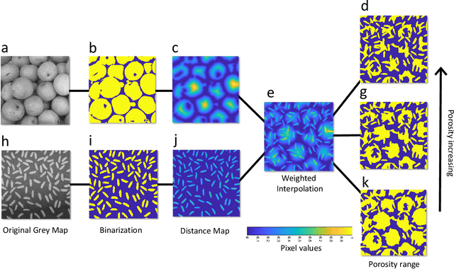
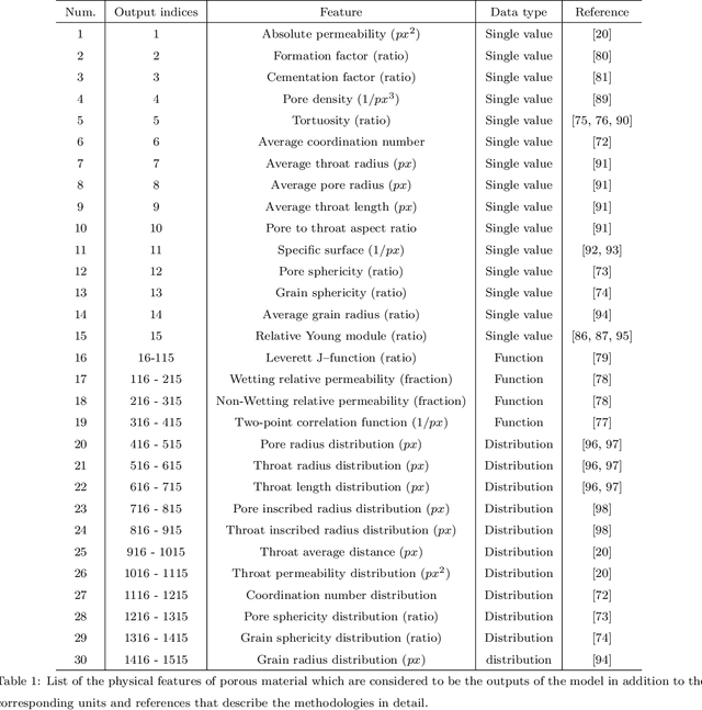
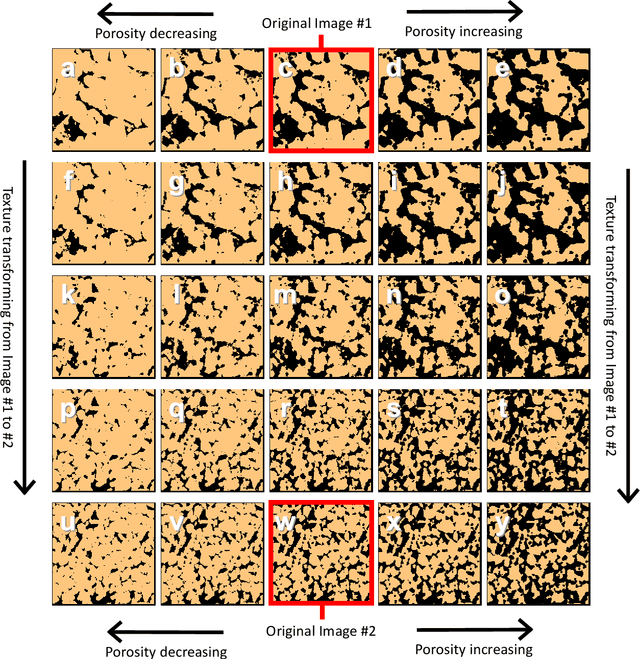
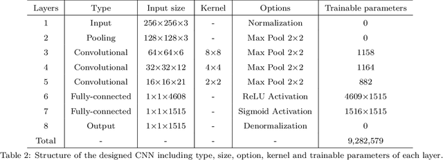
DeePore is a deep learning workflow for rapid estimation of a wide range of porous material properties based on the binarized micro-tomography images. We generated 17700 semi-real 3-D micro-structures of porous geo-materials and 30 physical properties of each sample are calculated using physical simulations on the corresponding pore network models. The dataset of porous material images obtained and physical features of them are unprecedented in terms of the number of samples and variety of the extracted features. Next, a re-designed feed-forward convolutional neural network is trained based on the dataset to estimate several morphological, hydraulic, electrical, and mechanical characteristics of porous material in a fraction of a second. The average coefficient of determination ($R^2$) for 3173 testing samples is 0.9385 which is very reasonable considering the wide range of micro-structure textures and extracted features. This workflow is compatible with any physical size of the images due to its dimensionless approach.