 Add to Chrome
 Add to Firefox
 Add to Edge
Applications of Epileptic Seizures Detection in Neuroimaging Modalities Using Deep Learning Techniques: Methods, Challenges, and Future Works
May 29, 2021
Epileptic seizures are a type of neurological disorder that affect many people worldwide. Specialist physicians and neurologists take advantage of structural and functional neuroimaging modalities to diagnose various types of epileptic seizures. Neuroimaging modalities assist specialist physicians considerably in analyzing brain tissue and the changes made in it. One method to accelerate the accurate and fast diagnosis of epileptic seizures is to employ computer aided diagnosis systems (CADS) based on artificial intelligence (AI) and functional and structural neuroimaging modalities. AI encompasses a variety of areas, and one of its branches is deep learning (DL). Not long ago, and before the rise of DL algorithms, feature extraction was an essential part of every conventional machine learning method, yet handcrafting features limit these models' performances to the knowledge of system designers. DL methods resolved this issue entirely by automating the feature extraction and classification process; applications of these methods in many fields of medicine, such as the diagnosis of epileptic seizures, have made notable improvements. In this paper, a comprehensive overview of the types of DL methods exploited to diagnose epileptic seizures from various neuroimaging modalities has been studied. Additionally, rehabilitation systems and cloud computing in epileptic seizures diagnosis applications have been exactly investigated using various modalities.
Applications of Deep Learning Techniques for Automated Multiple Sclerosis Detection Using Magnetic Resonance Imaging: A Review
May 11, 2021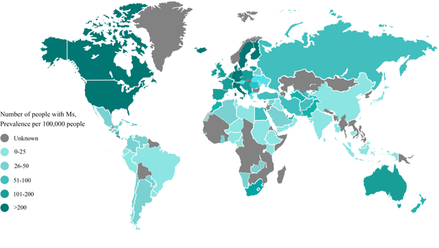
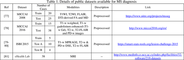
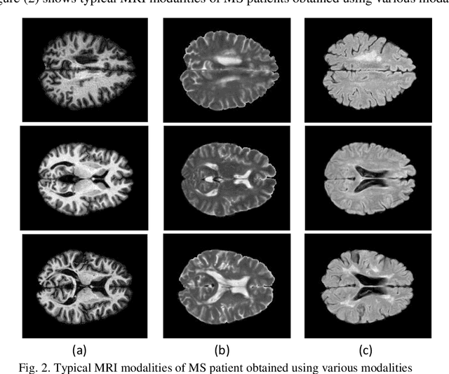
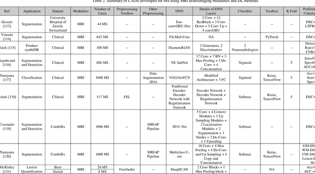
Multiple Sclerosis (MS) is a type of brain disease which causes visual, sensory, and motor problems for people with a detrimental effect on the functioning of the nervous system. In order to diagnose MS, multiple screening methods have been proposed so far; among them, magnetic resonance imaging (MRI) has received considerable attention among physicians. MRI modalities provide physicians with fundamental information about the structure and function of the brain, which is crucial for the rapid diagnosis of MS lesions. Diagnosing MS using MRI is time-consuming, tedious, and prone to manual errors. Hence, computer aided diagnosis systems (CADS) based on artificial intelligence (AI) methods have been proposed in recent years for accurate diagnosis of MS using MRI neuroimaging modalities. In the AI field, automated MS diagnosis is being conducted using (i) conventional machine learning and (ii) deep learning (DL) techniques. The conventional machine learning approach is based on feature extraction and selection by trial and error. In DL, these steps are performed by the DL model itself. In this paper, a complete review of automated MS diagnosis methods performed using DL techniques with MRI neuroimaging modalities are discussed. Also, each work is thoroughly reviewed and discussed. Finally, the most important challenges and future directions in the automated MS diagnosis using DL techniques coupled with MRI modalities are presented in detail.
Time Series Forecasting of New Cases and New Deaths Rate for COVID-19 using Deep Learning Methods
Apr 28, 2021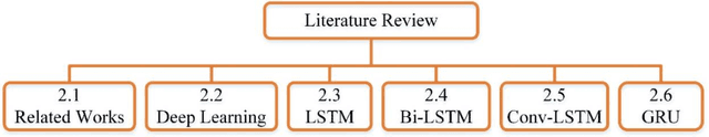
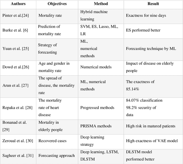
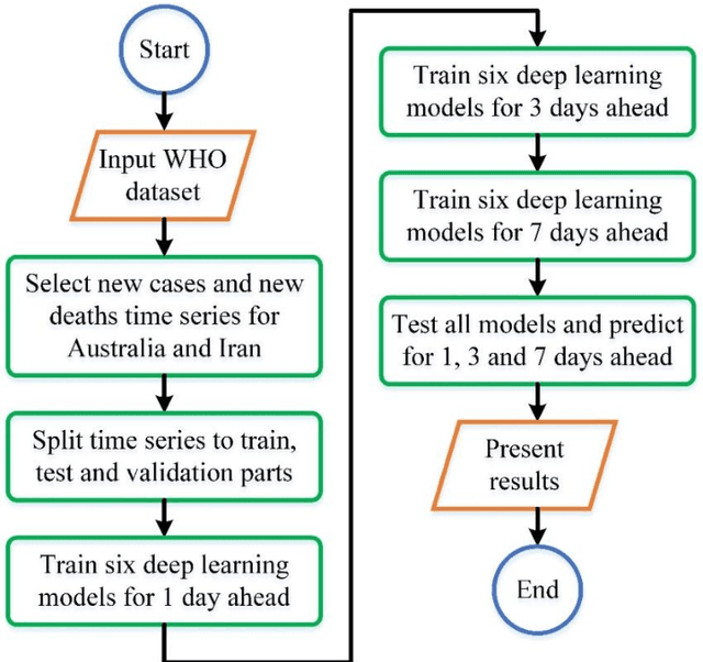
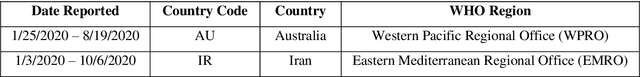
Covid-19 has been started in the year 2019 and imposed restrictions in many countries and costs organisations and governments. Predicting the number of new cases and deaths during this period can be a useful step in predicting the costs and facilities required in the future. The purpose of this study is to predict new cases and death rate for seven days ahead. Deep learning methods and statistical analysis model these predictions for 100 days. Six different deep learning methods are examined for the data adopted from the WHO website. Three methods are known as LSTM, Convolutional LSTM, and GRU. The bi-directional mode is then considered for each method to forecast the rate of new cases and new deaths for Australia and Iran countries. This study is novel as it attempts to implement the mentioned three deep learning methods, along with their Bi-directional models, to predict COVID-19 new cases and new death rate time series. All methods are compared, and results are presented. The results are examined in the form of graphs and statistical analyses. The results show that the Bi-directional models have lower error than other models. Several error evaluation metrics are presented to compare all models, and finally, the superiority of Bi-directional methods are determined. The experimental results and statistical test show on datasets to compare the proposed method with other baseline methods. This research could be useful for organisations working against COVID-19 and determining their long-term plans.
Automatic Diagnosis of COVID-19 from CT Images using CycleGAN and Transfer Learning
Apr 24, 2021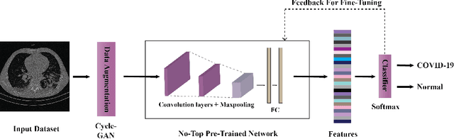
The outbreak of the corona virus disease (COVID-19) has changed the lives of most people on Earth. Given the high prevalence of this disease, its correct diagnosis in order to quarantine patients is of the utmost importance in steps of fighting this pandemic. Among the various modalities used for diagnosis, medical imaging, especially computed tomography (CT) imaging, has been the focus of many previous studies due to its accuracy and availability. In addition, automation of diagnostic methods can be of great help to physicians. In this paper, a method based on pre-trained deep neural networks is presented, which, by taking advantage of a cyclic generative adversarial net (CycleGAN) model for data augmentation, has reached state-of-the-art performance for the task at hand, i.e., 99.60% accuracy. Also, in order to evaluate the method, a dataset containing 3163 images from 189 patients has been collected and labeled by physicians. Unlike prior datasets, normal data have been collected from people suspected of having COVID-19 disease and not from data from other diseases, and this database is made available publicly.
CNN AE: Convolution Neural Network combined with Autoencoder approach to detect survival chance of COVID 19 patients
Apr 18, 2021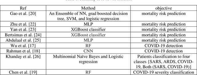
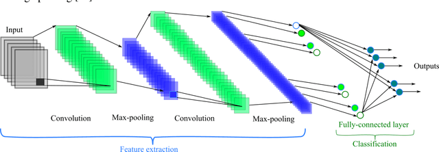
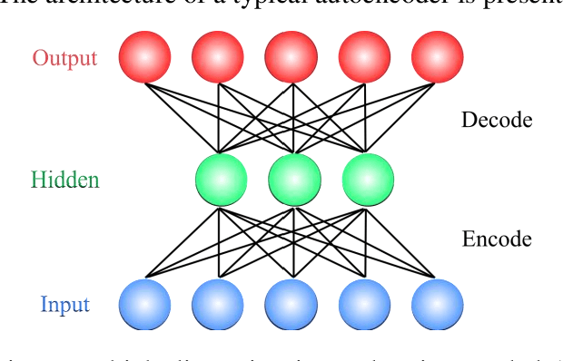
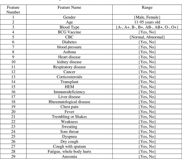
In this paper, we propose a novel method named CNN-AE to predict survival chance of COVID-19 patients using a CNN trained on clinical information. To further increase the prediction accuracy, we use the CNN in combination with an autoencoder. Our method is one of the first that aims to predict survival chance of already infected patients. We rely on clinical data to carry out the prediction. The motivation is that the required resources to prepare CT images are expensive and limited compared to the resources required to collect clinical data such as blood pressure, liver disease, etc. We evaluate our method on a publicly available clinical dataset of deceased and recovered patients which we have collected. Careful analysis of the dataset properties is also presented which consists of important features extraction and correlation computation between features. Since most of COVID-19 patients are usually recovered, the number of deceased samples of our dataset is low leading to data imbalance. To remedy this issue, a data augmentation procedure based on autoencoders is proposed. To demonstrate the generality of our augmentation method, we train random forest and Na\"ive Bayes on our dataset with and without augmentation and compare their performance. We also evaluate our method on another dataset for further generality verification. Experimental results reveal the superiority of CNN-AE method compared to the standard CNN as well as other methods such as random forest and Na\"ive Bayes. COVID-19 detection average accuracy of CNN-AE is 96.05% which is higher than CNN average accuracy of 92.49%. To show that clinical data can be used as a reliable dataset for COVID-19 survival chance prediction, CNN-AE is compared with a standard CNN which is trained on CT images.
An Overview on Artificial Intelligence Techniques for Diagnosis of Schizophrenia Based on Magnetic Resonance Imaging Modalities: Methods, Challenges, and Future Works
Feb 24, 2021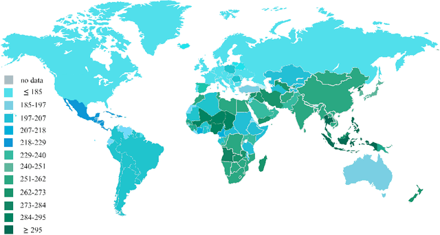
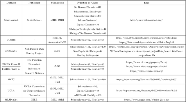
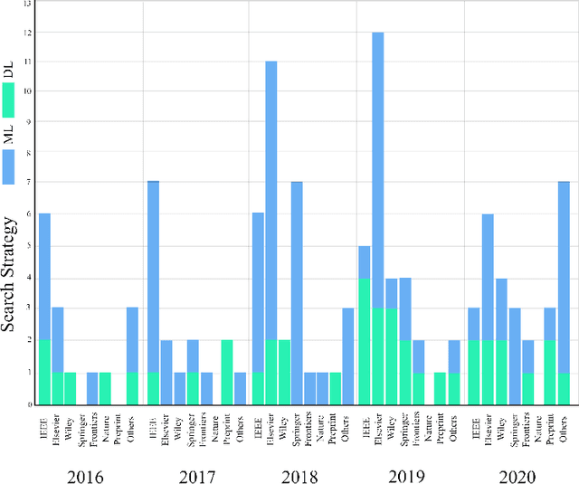
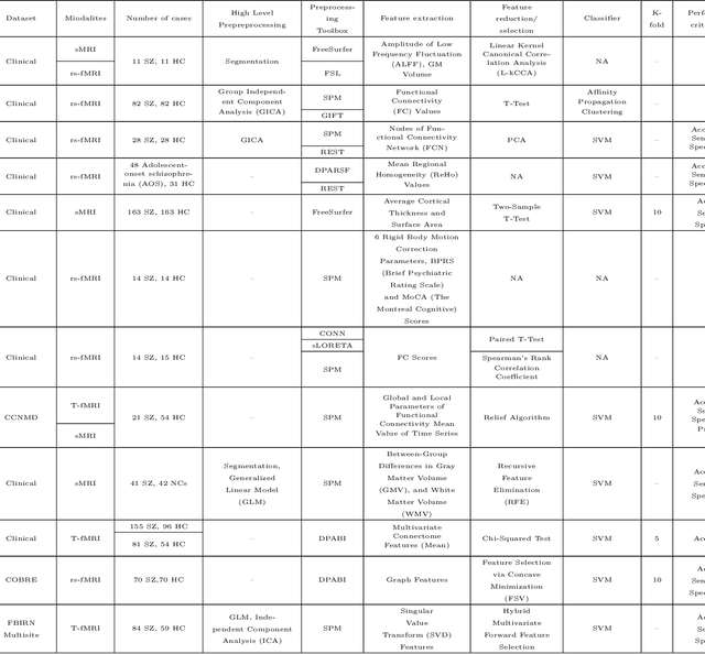
Schizophrenia (SZ) is a mental disorder that typically emerges in late adolescence or early adulthood. It reduces the life expectancy of patients by 15 years. Abnormal behavior, perception of emotions, social relationships, and reality perception are among its most significant symptoms. Past studies have revealed the temporal and anterior lobes of hippocampus regions of brain get affected by SZ. Also, increased volume of cerebrospinal fluid (CSF) and decreased volume of white and gray matter can be observed due to this disease. The magnetic resonance imaging (MRI) is the popular neuroimaging technique used to explore structural/functional brain abnormalities in SZ disorder owing to its high spatial resolution. Various artificial intelligence (AI) techniques have been employed with advanced image/signal processing methods to obtain accurate diagnosis of SZ. This paper presents a comprehensive overview of studies conducted on automated diagnosis of SZ using MRI modalities. Main findings, various challenges, and future works in developing the automated SZ detection are described in this paper.
Fusion of convolution neural network, support vector machine and Sobel filter for accurate detection of COVID-19 patients using X-ray images
Feb 13, 2021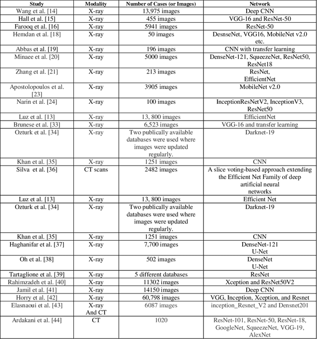
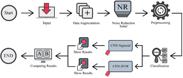
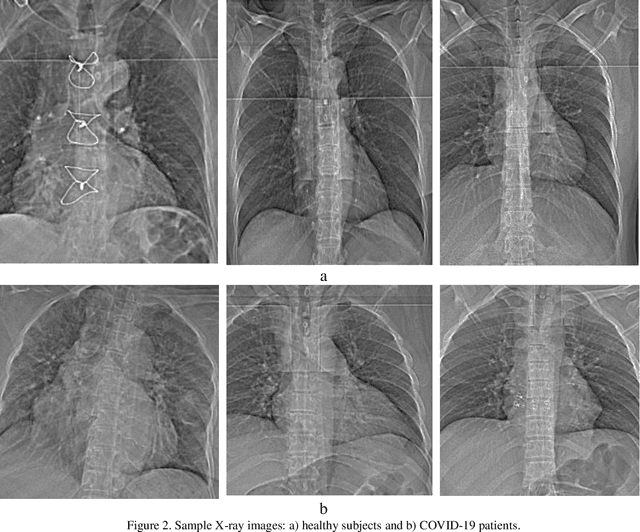
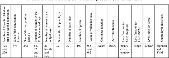
The coronavirus (COVID-19) is currently the most common contagious disease which is prevalent all over the world. The main challenge of this disease is the primary diagnosis to prevent secondary infections and its spread from one person to another. Therefore, it is essential to use an automatic diagnosis system along with clinical procedures for the rapid diagnosis of COVID-19 to prevent its spread. Artificial intelligence techniques using computed tomography (CT) images of the lungs and chest radiography have the potential to obtain high diagnostic performance for Covid-19 diagnosis. In this study, a fusion of convolutional neural network (CNN), support vector machine (SVM), and Sobel filter is proposed to detect COVID-19 using X-ray images. A new X-ray image dataset was collected and subjected to high pass filter using a Sobel filter to obtain the edges of the images. Then these images are fed to CNN deep learning model followed by SVM classifier with ten-fold cross validation strategy. This method is designed so that it can learn with not many data. Our results show that the proposed CNN-SVM with Sobel filtering (CNN-SVM+Sobel) achieved the highest classification accuracy of 99.02% in accurate detection of COVID-19. It showed that using Sobel filter can improve the performance of CNN. Unlike most of the other researches, this method does not use a pre-trained network. We have also validated our developed model using six public databases and obtained the highest performance. Hence, our developed model is ready for clinical application
Uncertainty-Aware Semi-supervised Method using Large Unlabelled and Limited Labeled COVID-19 Data
Feb 12, 2021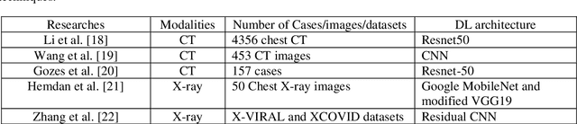
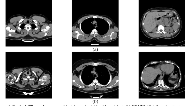
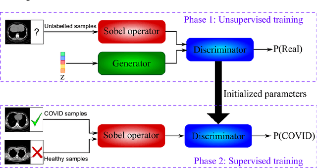
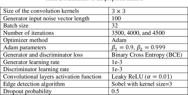
The new coronavirus has caused more than 1 million deaths and continues to spread rapidly. This virus targets the lungs, causing respiratory distress which can be mild or severe. The X-ray or computed tomography (CT) images of lungs can reveal whether the patient is infected with COVID-19 or not. Many researchers are trying to improve COVID-19 detection using artificial intelligence. In this paper, relying on Generative Adversarial Networks (GAN), we propose a Semi-supervised Classification using Limited Labelled Data (SCLLD) for automated COVID-19 detection. Our motivation is to develop learning method which can cope with scenarios that preparing labelled data is time consuming or expensive. We further improved the detection accuracy of the proposed method by applying Sobel edge detection. The GAN discriminator output is a probability value which is used for classification in this work. The proposed system is trained using 10,000 CT scans collected from Omid hospital. Also, we validate our system using the public dataset. The proposed method is compared with other state of the art supervised methods such as Gaussian processes. To the best of our knowledge, this is the first time a COVID-19 semi-supervised detection method is presented. Our method is capable of learning from a mixture of limited labelled and unlabelled data where supervised learners fail due to lack of sufficient amount of labelled data. Our semi-supervised training method significantly outperforms the supervised training of Convolutional Neural Network (CNN) in case labelled training data is scarce. Our method has achieved an accuracy of 99.60%, sensitivity of 99.39%, and specificity of 99.80% where CNN (trained supervised) has achieved an accuracy of 69.87%, sensitivity of 94%, and specificity of 46.40%.
Handling of uncertainty in medical data using machine learning and probability theory techniques: A review of 30 years
Aug 23, 2020
Understanding data and reaching valid conclusions are of paramount importance in the present era of big data. Machine learning and probability theory methods have widespread application for this purpose in different fields. One critically important yet less explored aspect is how data and model uncertainties are captured and analyzed. Proper quantification of uncertainty provides valuable information for optimal decision making. This paper reviewed related studies conducted in the last 30 years (from 1991 to 2020) in handling uncertainties in medical data using probability theory and machine learning techniques. Medical data is more prone to uncertainty due to the presence of noise in the data. So, it is very important to have clean medical data without any noise to get accurate diagnosis. The sources of noise in the medical data need to be known to address this issue. Based on the medical data obtained by the physician, diagnosis of disease, and treatment plan are prescribed. Hence, the uncertainty is growing in healthcare and there is limited knowledge to address these problems. We have little knowledge about the optimal treatment methods as there are many sources of uncertainty in medical science. Our findings indicate that there are few challenges to be addressed in handling the uncertainty in medical raw data and new models. In this work, we have summarized various methods employed to overcome this problem. Nowadays, application of novel deep learning techniques to deal such uncertainties have significantly increased.
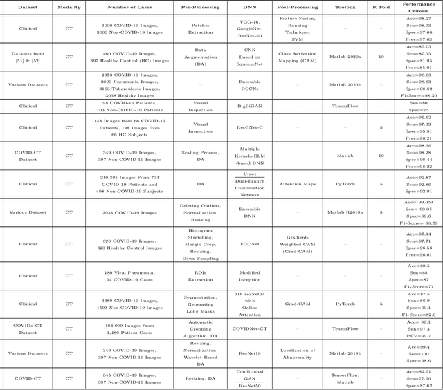
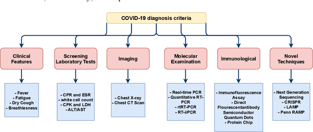
Automated Detection and Forecasting of COVID-19 using Deep Learning Techniques: A Review
Jul 27, 2020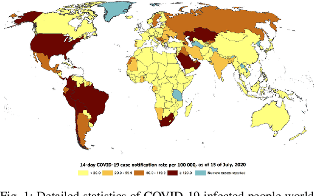
Coronavirus, or COVID-19, is a hazardous disease that has endangered the health of many people around the world by directly affecting the lungs. COVID-19 is a medium-sized, coated virus with a single-stranded RNA. This virus has one of the largest RNA genomes and is approximately 120 nm. The X-Ray and computed tomography (CT) imaging modalities are widely used to obtain a fast and accurate medical diagnosis. Identifying COVID-19 from these medical images is extremely challenging as it is time-consuming, demanding, and prone to human errors. Hence, artificial intelligence (AI) methodologies can be used to obtain consistent high performance. Among the AI methodologies, deep learning (DL) networks have gained much popularity compared to traditional machine learning (ML) methods. Unlike ML techniques, all stages of feature extraction, feature selection, and classification are accomplished automatically in DL models. In this paper, a complete survey of studies on the application of DL techniques for COVID-19 diagnostic and automated segmentation of lungs is discussed, concentrating on works that used X-Ray and CT images. Additionally, a review of papers on the forecasting of coronavirus prevalence in different parts of the world with DL techniques is presented. Lastly, the challenges faced in the automated detection of COVID-19 using DL techniques and directions for future research are discussed.