 Add to Chrome
 Add to Firefox
 Add to Edge
Compass: Prostate Cancer Detection Needs Multi-View Context
Jul 08, 2026
Artificial intelligence (AI) analysis of micro-ultrasound ($μ$US) has shown promise for prostate cancer (PCa) detection. However, most existing AI methods focus on the analysis of single $μ$US images in isolation. By contrast, expert $μ$US readers typically assess a full recorded video study, which provides three-dimensional context, to improve PCa detection compared to single-frame analysis. Inspired by this clinical workflow, we propose Compass, a novel AI methodology which models a $μ$US study as a stream of 2D images. Compass jointly integrates rotational sweep videos of the prostate with $μ$US frames acquired at the moment of biopsy, and performs evidence aggregation across the study using a transformer conditioned on the probe's rotational angle. Finally, a decoder head predicts frame-level and study-level risk scores for the patient. The model is trained and evaluated using a multi-center clinical trial dataset of $μ$US studies, including continuous rotational scans of the prostate and videos captured during biopsy acquisition. We compare the proposed method to baseline AI methods from the literature and to risk scores provided by clinical experts. Our framework shows strong performance, highlighting the value of multi-view context for $μ$US PCa detection, and providing a potentially powerful tool to complement human expertise in $μ$US-based PCa diagnosis. Our code is available at: https://github.com/mharmanani/Compass.

ProstNFound+: A Prospective Study using Medical Foundation Models for Prostate Cancer Detection
Oct 30, 2025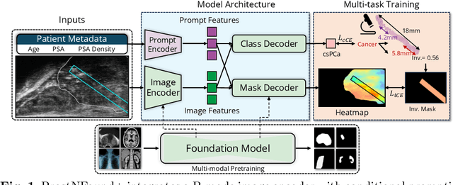
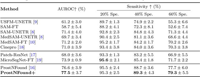
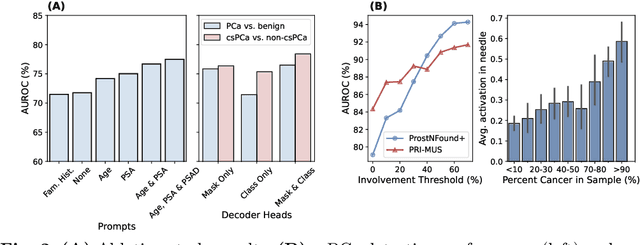
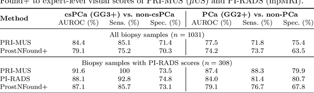
Purpose: Medical foundation models (FMs) offer a path to build high-performance diagnostic systems. However, their application to prostate cancer (PCa) detection from micro-ultrasound ({\mu}US) remains untested in clinical settings. We present ProstNFound+, an adaptation of FMs for PCa detection from {\mu}US, along with its first prospective validation. Methods: ProstNFound+ incorporates a medical FM, adapter tuning, and a custom prompt encoder that embeds PCa-specific clinical biomarkers. The model generates a cancer heatmap and a risk score for clinically significant PCa. Following training on multi-center retrospective data, the model is prospectively evaluated on data acquired five years later from a new clinical site. Model predictions are benchmarked against standard clinical scoring protocols (PRI-MUS and PI-RADS). Results: ProstNFound+ shows strong generalization to the prospective data, with no performance degradation compared to retrospective evaluation. It aligns closely with clinical scores and produces interpretable heatmaps consistent with biopsy-confirmed lesions. Conclusion: The results highlight its potential for clinical deployment, offering a scalable and interpretable alternative to expert-driven protocols.
Mask Enhanced Deeply Supervised Prostate Cancer Detection on B-mode Micro-Ultrasound
Dec 14, 2024


Prostate cancer is a leading cause of cancer-related deaths among men. The recent development of high frequency, micro-ultrasound imaging offers improved resolution compared to conventional ultrasound and potentially a better ability to differentiate clinically significant cancer from normal tissue. However, the features of prostate cancer remain subtle, with ambiguous borders with normal tissue and large variations in appearance, making it challenging for both machine learning and humans to localize it on micro-ultrasound images. We propose a novel Mask Enhanced Deeply-supervised Micro-US network, termed MedMusNet, to automatically and more accurately segment prostate cancer to be used as potential targets for biopsy procedures. MedMusNet leverages predicted masks of prostate cancer to enforce the learned features layer-wisely within the network, reducing the influence of noise and improving overall consistency across frames. MedMusNet successfully detected 76% of clinically significant cancer with a Dice Similarity Coefficient of 0.365, significantly outperforming the baseline Swin-M2F in specificity and accuracy (Wilcoxon test, Bonferroni correction, p-value<0.05). While the lesion-level and patient-level analyses showed improved performance compared to human experts and different baseline, the improvements did not reach statistical significance, likely on account of the small cohort. We have presented a novel approach to automatically detect and segment clinically significant prostate cancer on B-mode micro-ultrasound images. Our MedMusNet model outperformed other models, surpassing even human experts. These preliminary results suggest the potential for aiding urologists in prostate cancer diagnosis via biopsy and treatment decision-making.