 Add to Chrome
 Add to Firefox
 Add to Edge
Mass Segmentation From Mammograms
Papers and Code
Semi- and Weakly-Supervised Learning for Mammogram Mass Segmentation with Limited Annotations
Mar 14, 2024


Accurate identification of breast masses is crucial in diagnosing breast cancer; however, it can be challenging due to their small size and being camouflaged in surrounding normal glands. Worse still, it is also expensive in clinical practice to obtain adequate pixel-wise annotations for training deep neural networks. To overcome these two difficulties with one stone, we propose a semi- and weakly-supervised learning framework for mass segmentation that utilizes limited strongly-labeled samples and sufficient weakly-labeled samples to achieve satisfactory performance. The framework consists of an auxiliary branch to exclude lesion-irrelevant background areas, a segmentation branch for final prediction, and a spatial prompting module to integrate the complementary information of the two branches. We further disentangle encoded obscure features into lesion-related and others to boost performance. Experiments on CBIS-DDSM and INbreast datasets demonstrate the effectiveness of our method.
Intelligent Breast Cancer Diagnosis with Heuristic-assisted Trans-Res-U-Net and Multiscale DenseNet using Mammogram Images
Oct 30, 2023Breast cancer (BC) significantly contributes to cancer-related mortality in women, underscoring the criticality of early detection for optimal patient outcomes. A mammography is a key tool for identifying and diagnosing breast abnormalities; however, accurately distinguishing malignant mass lesions remains challenging. To address this issue, we propose a novel deep learning approach for BC screening utilizing mammography images. Our proposed model comprises three distinct stages: data collection from established benchmark sources, image segmentation employing an Atrous Convolution-based Attentive and Adaptive Trans-Res-UNet (ACA-ATRUNet) architecture, and BC identification via an Atrous Convolution-based Attentive and Adaptive Multi-scale DenseNet (ACA-AMDN) model. The hyperparameters within the ACA-ATRUNet and ACA-AMDN models are optimised using the Modified Mussel Length-based Eurasian Oystercatcher Optimization (MML-EOO) algorithm. Performance evaluation, leveraging multiple metrics, is conducted, and a comparative analysis against conventional methods is presented. Our experimental findings reveal that the proposed BC detection framework attains superior precision rates in early disease detection, demonstrating its potential to enhance mammography-based screening methodologies.
Mammograms Classification: A Review
Mar 04, 2022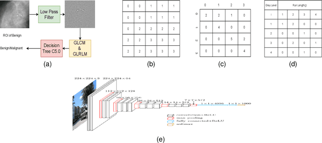
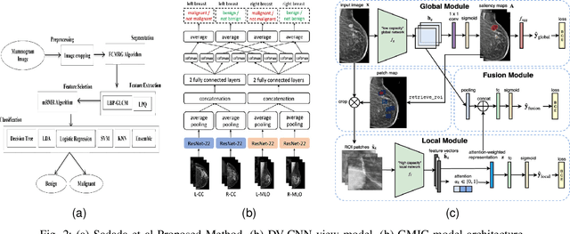
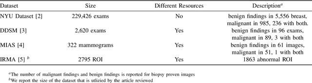
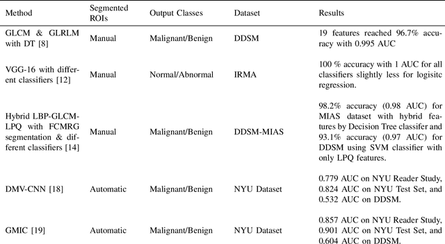
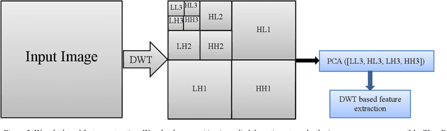
An advanced reliable low-cost form of screening method, Digital mammography has been used as an effective imaging method for breast cancer detection. With an increased focus on technologies to aid healthcare, Mammogram images have been utilized in developing computer-aided diagnosis systems that will potentially help in clinical diagnosis. Researchers have proved that artificial intelligence with its emerging technologies can be used in the early detection of the disease and improve radiologists' performance in assessing breast cancer. In this paper, we review the methods developed for mammogram mass classification in two categories. The first one is classifying manually provided cropped region of interests (ROI) as either malignant or benign, and the second one is the classification of automatically segmented ROIs as either malignant or benign. We also provide an overview of datasets and evaluation metrics used in the classification task. Finally, we compare and discuss the deep learning approach to classical image processing and learning approach in this domain.
Leveraging SLIC Superpixel Segmentation and Cascaded Ensemble SVM for Fully Automated Mass Detection In Mammograms
Oct 20, 2020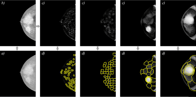
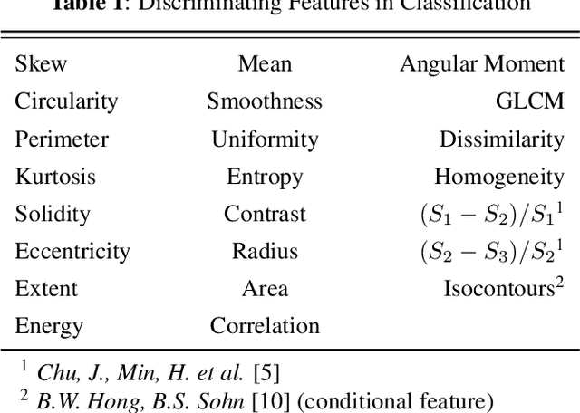
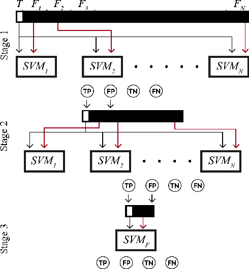
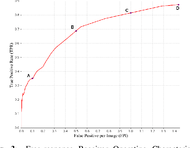
Identification and segmentation of breast masses in mammograms face complex challenges, owing to the highly variable nature of malignant densities with regards to their shape, contours, texture and orientation. Additionally, classifiers typically suffer from high class imbalance in region candidates, where normal tissue regions vastly outnumber malignant masses. This paper proposes a rigorous segmentation method, supported by morphological enhancement using grayscale linear filters. A novel cascaded ensemble of support vector machines (SVM) is used to effectively tackle the class imbalance and provide significant predictions. For True Positive Rate (TPR) of 0.35, 0.69 and 0.82, the system generates only 0.1, 0.5 and 1.0 False Positives/Image (FPI), respectively.
Dual Convolutional Neural Networks for Breast Mass Segmentation and Diagnosis in Mammography
Aug 11, 2020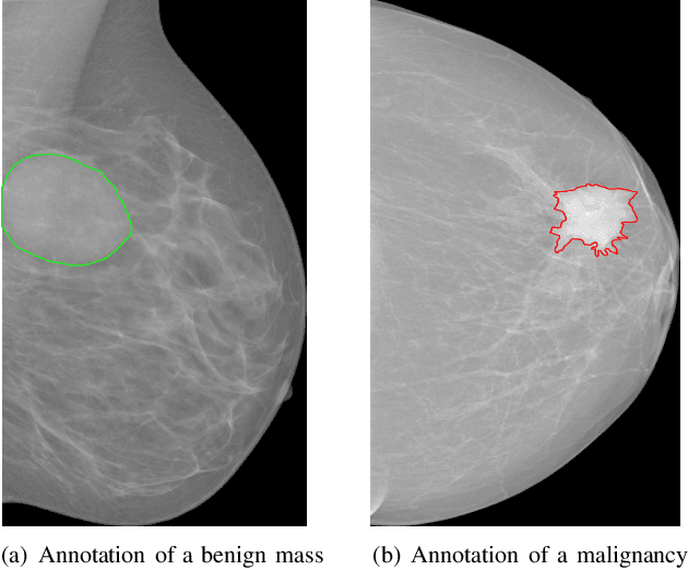
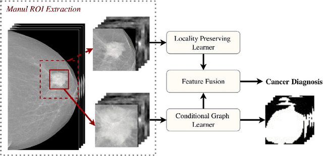
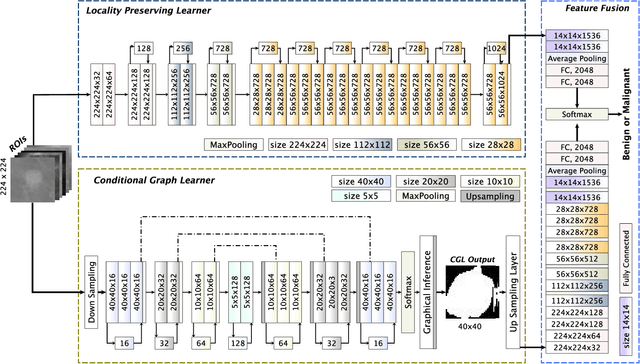
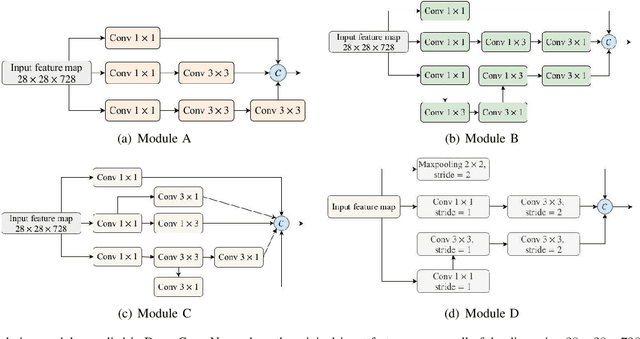
Deep convolutional neural networks (CNNs) have emerged as a new paradigm for Mammogram diagnosis. Contemporary CNN-based computer-aided-diagnosis (CAD) for breast cancer directly extract latent features from input mammogram image and ignore the importance of morphological features. In this paper, we introduce a novel deep learning framework for mammogram image processing, which computes mass segmentation and simultaneously predict diagnosis results. Specifically, our method is constructed in a dual-path architecture that solves the mapping in a dual-problem manner, with an additional consideration of important shape and boundary knowledge. One path called the Locality Preserving Learner (LPL), is devoted to hierarchically extracting and exploiting intrinsic features of the input. Whereas the other path, called the Conditional Graph Learner (CGL) focuses on generating geometrical features via modeling pixel-wise image to mask correlations. By integrating the two learners, both the semantics and structure are well preserved and the component learning paths in return complement each other, contributing an improvement to the mass segmentation and cancer classification problem at the same time. We evaluated our method on two most used public mammography datasets, DDSM and INbreast. Experimental results show that DualCoreNet achieves the best mammography segmentation and classification simultaneously, outperforming recent state-of-the-art models.
Two-stage breast mass detection and segmentation system towards automated high-resolution full mammogram analysis
Feb 27, 2020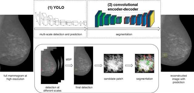
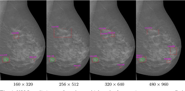
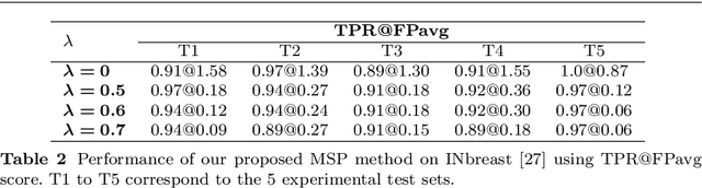
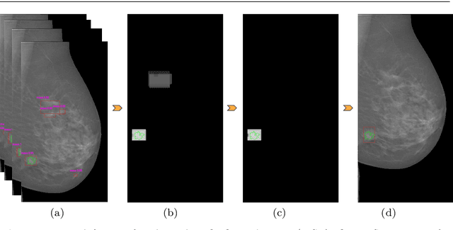
Mammography is the primary imaging modality used for early detection and diagnosis of breast cancer. Mammography analysis mainly refers to the extraction of regions of interest around tumors, followed by a segmentation step, which is essential to further classification of benign or malignant tumors. Breast masses are the most important findings among breast abnormalities. However, manual delineation of masses from native mammogram is a time consuming and error-prone task. An integrated computer-aided diagnosis system to assist radiologists in automatically detecting and segmenting breast masses is therefore in urgent need. We propose a fully-automated approach that guides accurate mass segmentation from full mammograms at high resolution through a detection stage. First, mass detection is performed by an efficient deep learning approach, You-Only-Look-Once, extended by integrating multi-scale predictions to improve automatic candidate selection. Second, a convolutional encoder-decoder network using nested and dense skip connections is employed to fine-delineate candidate masses. Unlike most previous studies based on segmentation from regions, our framework handles mass segmentation from native full mammograms without user intervention. Trained on INbreast and DDSM-CBIS public datasets, the pipeline achieves an overall average Dice of 80.44% on high-resolution INbreast test images, outperforming state-of-the-art methods. Our system shows promising accuracy as an automatic full-image mass segmentation system. The comprehensive evaluation provided for both detection and segmentation stages reveals strong robustness to the diversity of size, shape and appearance of breast masses, towards better computer-aided diagnosis.
Applying a random projection algorithm to optimize machine learning model for breast lesion classification
Sep 09, 2020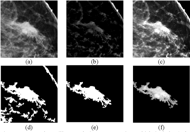
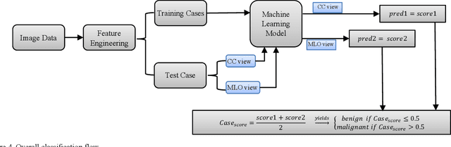
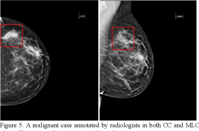
Machine learning is widely used in developing computer-aided diagnosis (CAD) schemes of medical images. However, CAD usually computes large number of image features from the targeted regions, which creates a challenge of how to identify a small and optimal feature vector to build robust machine learning models. In this study, we investigate feasibility of applying a random projection algorithm to build an optimal feature vector from the initially CAD-generated large feature pool and improve performance of machine learning model. We assemble a retrospective dataset involving 1,487 cases of mammograms in which 644 cases have confirmed malignant mass lesions and 843 have benign lesions. A CAD scheme is first applied to segment mass regions and initially compute 181 features. Then, support vector machine (SVM) models embedded with several feature dimensionality reduction methods are built to predict likelihood of lesions being malignant. All SVM models are trained and tested using a leave-one-case-out cross-validation method. SVM generates a likelihood score of each segmented mass region depicting on one-view mammogram. By fusion of two scores of the same mass depicting on two-view mammograms, a case-based likelihood score is also evaluated. Comparing with the principle component analyses, nonnegative matrix factorization, and Chi-squared methods, SVM embedded with the random projection algorithm yielded a significantly higher case-based lesion classification performance with the area under ROC curve of 0.84+0.01 (p<0.02). The study demonstrates that the random project algorithm is a promising method to generate optimal feature vectors to help improve performance of machine learning models of medical images.
Fully automatic computer-aided mass detection and segmentation via pseudo-color mammograms and Mask R-CNN
Jun 28, 2019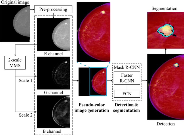
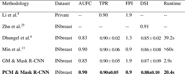
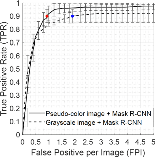
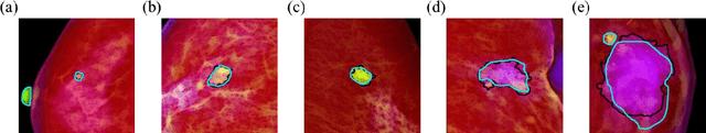
Purpose: To propose pseudo-color mammograms that enhance mammographic masses as part of a fast computer-aided detection (CAD) system that simultaneously detects and segments masses without any user intervention. Methods: The proposed pseudo-color mammograms, whose three channels contain the original grayscale mammogram and two morphologically enhanced images, are used to provide pseudo-color contrast to the lesions. The morphological enhancement 'sifts' out the mass-like mammographic patterns to improve detection and segmentation. We construct a fast, fully automated simultaneous mass detection and segmentation CAD system using the colored mammograms as inputs of transfer learning with the Mask R-CNN which is a state-of-the-art deep learning framework. The source code for this work has been made available online. Results: Evaluated on the publicly available mammographic dataset INbreast, the method outperforms the state-of-the-art methods by achieving an average true positive rate of 0.90 at 0.9 false positive per image and an average Dice similarity index for mass segmentation of 0.88, while taking 20.4 seconds to process each image on average. Conclusions: The proposed method provides an accurate, fully-automatic breast mass detection and segmentation result in less than half a minute without any user intervention while outperforming state-of-the-art methods.
Improved Breast Mass Segmentation in Mammograms with Conditional Residual U-net
Aug 27, 2018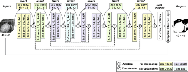
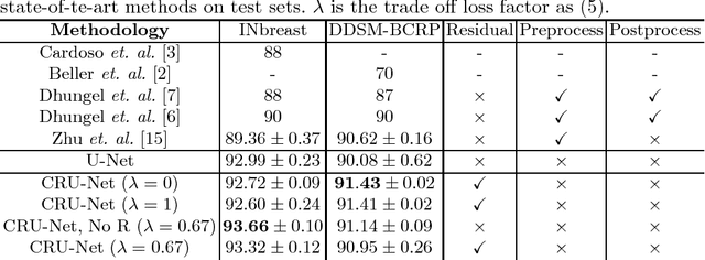
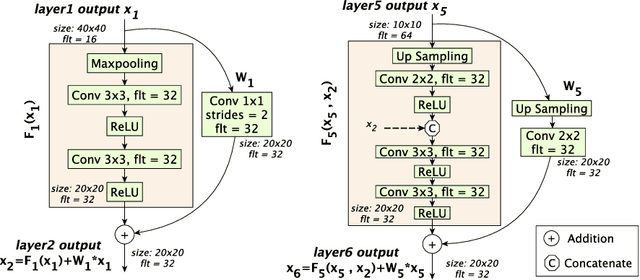
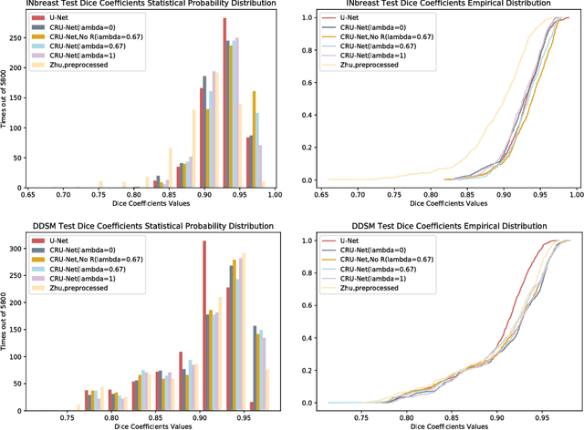
We explore the use of deep learning for breast mass segmentation in mammograms. By integrating the merits of residual learning and probabilistic graphical modelling with standard U-Net, we propose a new deep network, Conditional Residual U-Net (CRU-Net), to improve the U-Net segmentation performance. Benefiting from the advantage of probabilistic graphical modelling in the pixel-level labelling, and the structure insights of a deep residual network in the feature extraction, the CRU-Net provides excellent mass segmentation performance. Evaluations based on INbreast and DDSM-BCRP datasets demonstrate that the CRU-Net achieves the best mass segmentation performance compared to the state-of-art methodologies. Moreover, neither tedious pre-processing nor post-processing techniques are not required in our algorithm.
Deep Learning for Automated Medical Image Analysis
Mar 12, 2019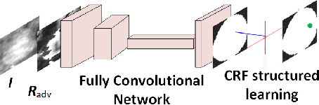
Medical imaging is an essential tool in many areas of medical applications, used for both diagnosis and treatment. However, reading medical images and making diagnosis or treatment recommendations require specially trained medical specialists. The current practice of reading medical images is labor-intensive, time-consuming, costly, and error-prone. It would be more desirable to have a computer-aided system that can automatically make diagnosis and treatment recommendations. Recent advances in deep learning enable us to rethink the ways of clinician diagnosis based on medical images. In this thesis, we will introduce 1) mammograms for detecting breast cancers, the most frequently diagnosed solid cancer for U.S. women, 2) lung CT images for detecting lung cancers, the most frequently diagnosed malignant cancer, and 3) head and neck CT images for automated delineation of organs at risk in radiotherapy. First, we will show how to employ the adversarial concept to generate the hard examples improving mammogram mass segmentation. Second, we will demonstrate how to use the weakly labeled data for the mammogram breast cancer diagnosis by efficiently design deep learning for multi-instance learning. Third, the thesis will walk through DeepLung system which combines deep 3D ConvNets and GBM for automated lung nodule detection and classification. Fourth, we will show how to use weakly labeled data to improve existing lung nodule detection system by integrating deep learning with a probabilistic graphic model. Lastly, we will demonstrate the AnatomyNet which is thousands of times faster and more accurate than previous methods on automated anatomy segmentation.