 Add to Chrome
 Add to Firefox
 Add to Edge
A Mobile App for Wound Localization using Deep Learning
Sep 15, 2020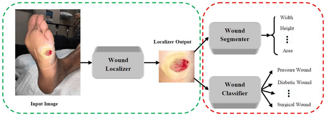
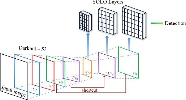
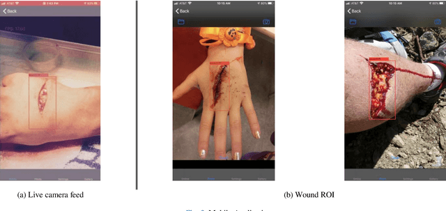
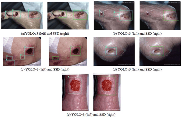
We present an automated wound localizer from 2D wound and ulcer images by using deep neural network, as the first step towards building an automated and complete wound diagnostic system. The wound localizer has been developed by using YOLOv3 model, which is then turned into an iOS mobile application. The developed localizer can detect the wound and its surrounding tissues and isolate the localized wounded region from images, which would be very helpful for future processing such as wound segmentation and classification due to the removal of unnecessary regions from wound images. For Mobile App development with video processing, a lighter version of YOLOv3 named tiny-YOLOv3 has been used. The model is trained and tested on our own image dataset in collaboration with AZH Wound and Vascular Center, Milwaukee, Wisconsin. The YOLOv3 model is compared with SSD model, showing that YOLOv3 gives a mAP value of 93.9%, which is much better than the SSD model (86.4%). The robustness and reliability of these models are also tested on a publicly available dataset named Medetec and shows a very good performance as well.
An Application of Manifold Learning in Global Shape Descriptors
Jan 08, 2019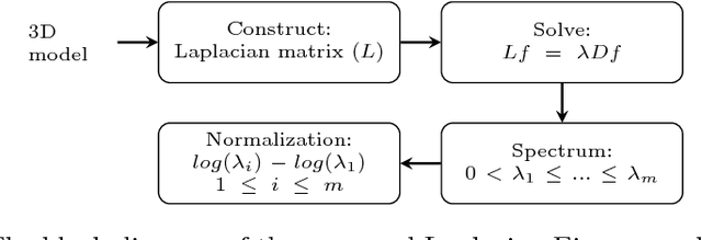
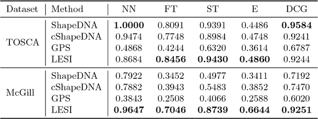
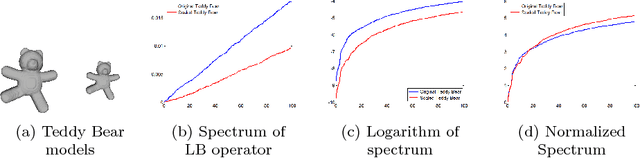

With the rapid expansion of applied 3D computational vision, shape descriptors have become increasingly important for a wide variety of applications and objects from molecules to planets. Appropriate shape descriptors are critical for accurate (and efficient) shape retrieval and 3D model classification. Several spectral-based shape descriptors have been introduced by solving various physical equations over a 3D surface model. In this paper, for the first time, we incorporate a specific group of techniques in statistics and machine learning, known as manifold learning, to develop a global shape descriptor in the computer graphics domain. The proposed descriptor utilizes the Laplacian Eigenmap technique in which the Laplacian eigenvalue problem is discretized using an exponential weighting scheme. As a result, our descriptor eliminates the limitations tied to the existing spectral descriptors, namely dependency on triangular mesh representation and high intra-class quality of 3D models. We also present a straightforward normalization method to obtain a scale-invariant descriptor. The extensive experiments performed in this study show that the present contribution provides a highly discriminative and robust shape descriptor under the presence of a high level of noise, random scale variations, and low sampling rate, in addition to the known isometric-invariance property of the Laplace-Beltrami operator. The proposed method significantly outperforms state-of-the-art algorithms on several non-rigid shape retrieval benchmarks.
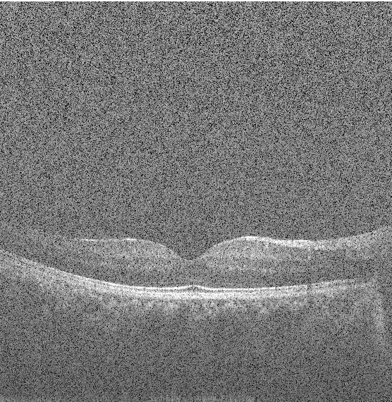
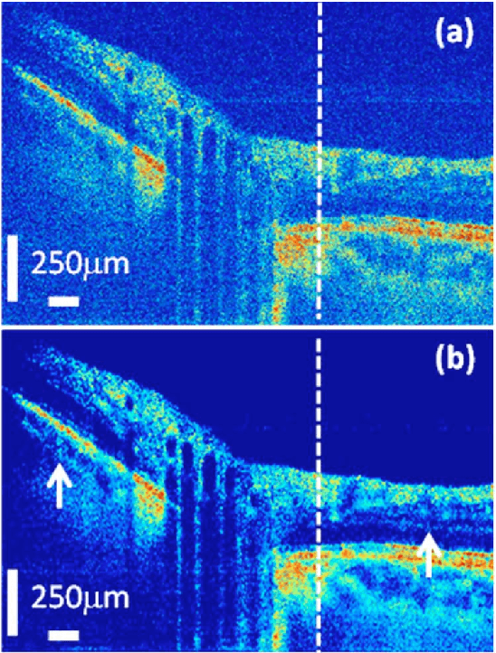
Application of Independent Component Analysis Techniques in Speckle Noise Reduction of Retinal OCT Images
Jul 28, 2015
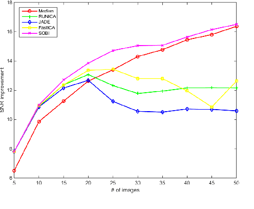
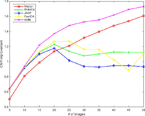
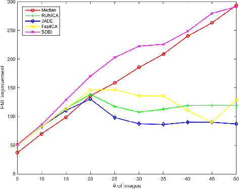
Optical Coherence Tomography (OCT) is an emerging technique in the field of biomedical imaging, with applications in ophthalmology, dermatology, coronary imaging etc. OCT images usually suffer from a granular pattern, called speckle noise, which restricts the process of interpretation. Therefore the need for speckle noise reduction techniques is of high importance. To the best of our knowledge, use of Independent Component Analysis (ICA) techniques has never been explored for speckle reduction of OCT images. Here, a comparative study of several ICA techniques (InfoMax, JADE, FastICA and SOBI) is provided for noise reduction of retinal OCT images. Having multiple B-scans of the same location, the eye movements are compensated using a rigid registration technique. Then, different ICA techniques are applied to the aggregated set of B-scans for extracting the noise-free image. Signal-to-Noise-Ratio (SNR), Contrast-to-Noise-Ratio (CNR) and Equivalent-Number-of-Looks (ENL), as well as analysis on the computational complexity of the methods, are considered as metrics for comparison. The results show that use of ICA can be beneficial, especially in case of having fewer number of B-scans.
siftservice.com - Turning a Computer Vision algorithm into a World Wide Web Service
Apr 11, 2015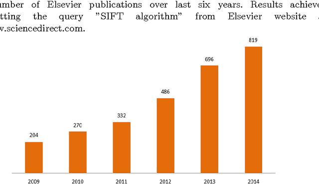
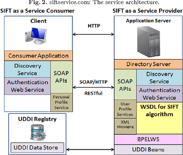
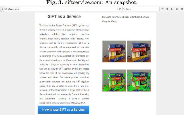
Image features detection and description is a longstanding topic in computer vision and pattern recognition areas. The Scale Invariant Feature Transform (SIFT) is probably the most popular and widely demanded feature descriptor which facilitates a variety of computer vision applications such as image registration, object tracking, image forgery detection, and 3D surface reconstruction. This work introduces a Software as a Service (SaaS) based implementation of the SIFT algorithm which is freely available at http://siftservice.com for any academic, educational and research purposes. The service provides application-to-application interaction and aims Rapid Application Development (RAD) and also fast prototyping for computer vision students and researchers all around the world. An Internet connection is all they need!
Sparse And Low Rank Decomposition Based Batch Image Alignment for Speckle Reduction of retinal OCT Images
Feb 09, 2015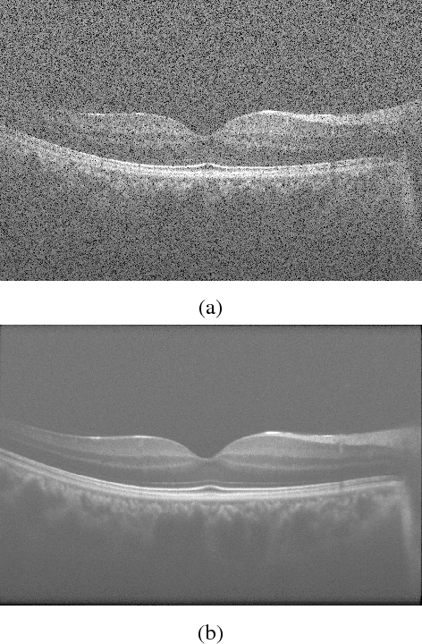
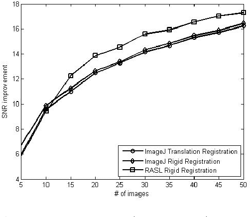
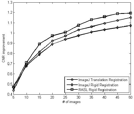
Optical Coherence Tomography (OCT) is an emerging technique in the field of biomedical imaging, with applications in ophthalmology, dermatology, coronary imaging etc. Due to the underlying physics, OCT images usually suffer from a granular pattern, called speckle noise, which restricts the process of interpretation. Here, a sparse and low rank decomposition based method is used for speckle reduction in retinal OCT images. This technique works on input data that consists of several B-scans of the same location. The next step is the batch alignment of the images using a sparse and low-rank decomposition based technique. Finally the denoised image is created by median filtering of the low-rank component of the processed data. Simultaneous decomposition and alignment of the images result in better performance in comparison to simple registration-based methods that are used in the literature for noise reduction of OCT images.
Structure Tensor Based Image Interpolation Method
Dec 26, 2014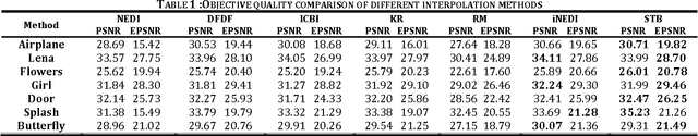
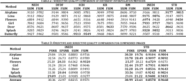

Feature preserving image interpolation is an active area in image processing field. In this paper a new direct edge directed image super-resolution algorithm based on structure tensors is proposed. Using an isotropic Gaussian filter, the structure tensor at each pixel of the input image is computed and the pixels are classified to three distinct classes; uniform region, corners and edges, according to the eigenvalues of the structure tensor. Due to application of the isotropic Gaussian filter, the classification is robust to noise presented in image. Based on the tangent eigenvector of the structure tensor, the edge direction is determined and used for interpolation along the edges. In comparison to some previous edge directed image interpolation methods, the proposed method achieves higher quality in both subjective and objective aspects. Also the proposed method outperforms previous methods in case of noisy and JPEG compressed images. Furthermore, without the need for optimization in the process, the algorithm can achieve higher speed.
State-of-the-Art in Retinal Optical Coherence Tomography Image Analysis
Nov 17, 2014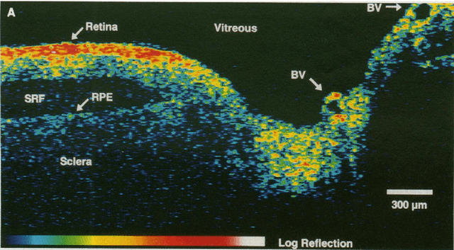
Optical Coherence Tomography (OCT) is one of the most emerging imaging modalities that has been used widely in the field of biomedical imaging. From its emergence in 1990's, plenty of hardware and software improvements have been made. Its applications range from ophthalmology to dermatology to coronary imaging etc. Here, the focus is on applications of OCT in ophthalmology and retinal imaging. OCT is able to non-invasively produce cross-sectional volume images of the tissues which are further used for analysis of the tissue structure and its properties. Due to the underlying physics, OCT images usually suffer from a granular pattern, called speckle noise, which restricts the process of interpretation, hence requiring specialized noise reduction techniques to remove the noise while preserving image details. Also, given the fact that OCT images are in the $\mu m$ -level, further analysis in needed to distinguish between the different structures in the imaged volume. Therefore the use of different segmentation techniques are of high importance. The movement of the tissue under imaging or the progression of disease in the tissue also imposes further implications both on the quality and the proper interpretation of the acquired images. Thus, use of image registration techniques can be very helpful. In this work, an overview of such image analysis techniques will be given.
Fast Mesh-Based Medical Image Registration
Nov 08, 2014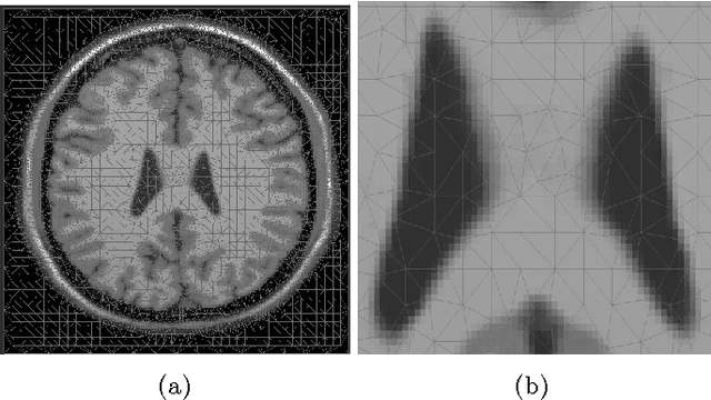
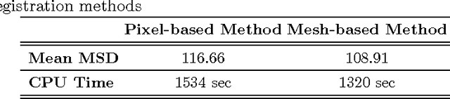
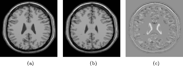
In this paper a fast triangular mesh based registration method is proposed. Having Template and Reference images as inputs, the template image is triangulated using a content adaptive mesh generation algorithm. Considering the pixel values at mesh nodes, interpolated using spline interpolation method for both of the images, the energy functional needed for image registration is minimized. The minimization process was achieved using a mesh based discretization of the distance measure and regularization term which resulted in a sparse system of linear equations, which due to the smaller size in comparison to the pixel-wise registration method, can be solved directly. Mean Squared Difference (MSD) is used as a metric for evaluating the results. Using the mesh based technique, higher speed was achieved compared to pixel-based curvature registration technique with fast DCT solver. The implementation was done in MATLAB without any specific optimization. Higher speeds can be achieved using C/C++ implementations.
Adaptive Mesh Representation and Restoration of Biomedical Images
Jun 27, 2014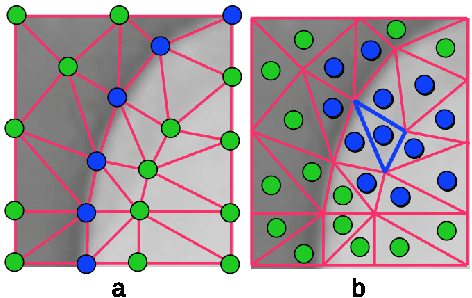
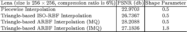
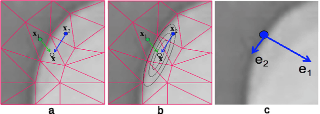
The triangulation of images has become an active research area in recent years for its compressive representation and ease of image processing and visualization. However, little work has been done on how to faithfully recover image intensities from a triangulated mesh of an image, a process also known as image restoration or decoding from meshes. The existing methods such as linear interpolation, least-square interpolation, or interpolation based on radial basis functions (RBFs) work to some extent, but often yield blurred features (edges, corners, etc.). The main reason for this problem is due to the isotropically-defined Euclidean distance that is taken into consideration in these methods, without considering the anisotropicity of feature intensities in an image. Moreover, most existing methods use intensities defined at mesh nodes whose intensities are often ambiguously defined on or near image edges (or feature boundaries). In the current paper, a new method of restoring an image from its triangulation representation is proposed, by utilizing anisotropic radial basis functions (ARBFs). This method considers not only the geometrical (Euclidean) distances but also the local feature orientations (anisotropic intensities). Additionally, this method is based on the intensities of mesh faces instead of mesh nodes and thus provides a more robust restoration. The two strategies together guarantee excellent feature-preserving restoration of an image with arbitrary super-resolutions from its triangulation representation, as demonstrated by various experiments provided in the paper.
An Optimization Method For Slice Interpolation Of Medical Images
Mar 27, 2014
Slice interpolation is a fast growing field in medical image processing. Intensity-based interpolation and object-based interpolation are two major groups of methods in the literature. In this paper, we describe an object-oriented, optimization method based on a modified version of curvature-based image registration, in which a displacement field is computed for the missing slice between two known slices and used to interpolate the intensities of the missing slice. The proposed approach is evaluated quantitatively by using the Mean Squared Difference (MSD) as a metric. The produced results also show visual improvement in preserving sharp edges in images.