 Add to Chrome
 Add to Firefox
 Add to Edge
Robust Principal Component Analysis for Background Estimation of Particle Image Velocimetry Data
Aug 16, 2019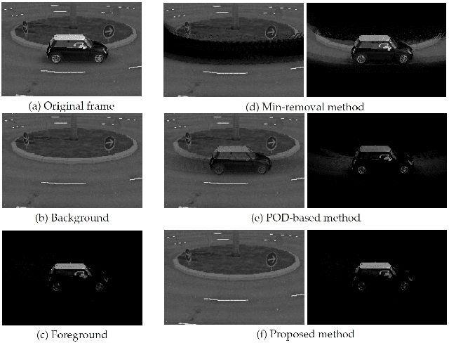
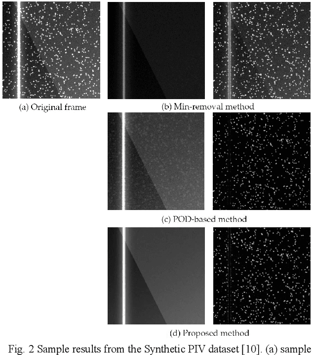
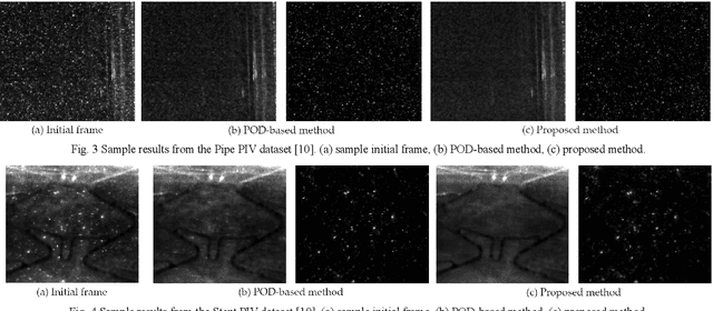
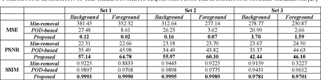
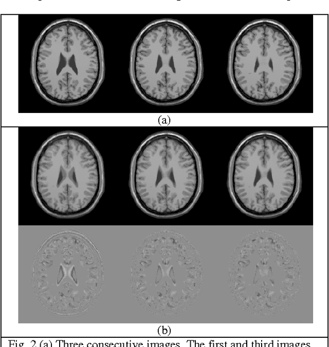
Particle Image Velocimetry (PIV) data processing procedures are adversely affected by light reflections and backgrounds as well as defects in the models and sticky particles that occlude the inner walls of the boundaries. In this paper, a novel approach is proposed for decomposition of the PIV data into background/foreground components, greatly reducing the effects of such artifacts. This is achieved by utilizing Robust Principal Component Analysis (RPCA) applied to the data matrix, generated by aggregating the vectorized PIV frames. It is assumed that the data matrix can be decomposed into two statistically different components, a low-rank component depicting the still background and a sparse component representing the moving particles within the imaged geometry. Formulating the assumptions as an optimization problem, Augmented Lagrange Multiplier (ALM) method is used for decomposing the data matrix into the low-rank and sparse components. Experiments and comparisons with the state-of-the-art using several PIV image sequences reveal the superiority of the proposed approach for background removal of PIV data.
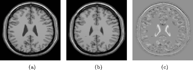
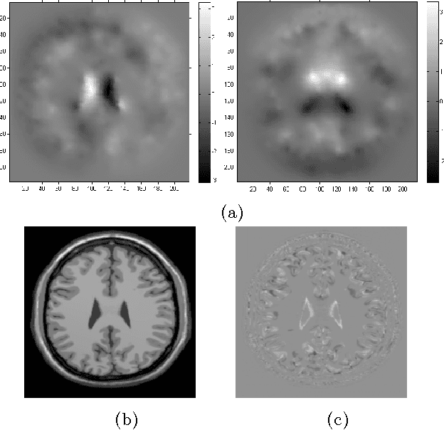
Markov Random Field Model-Based Salt and Pepper Noise Removal
Sep 20, 2016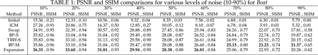
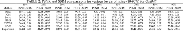
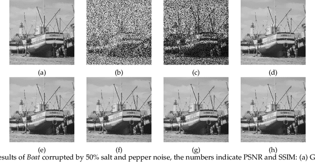
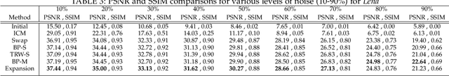
Problem of impulse noise reduction is a very well studied problem in image processing community and many different approaches have been proposed to tackle this problem. In the current work, the problem of fixed value impulse noise (salt and pepper) removal from images is investigated by use of a Markov Random Field (MRF) models with smoothness priors. After the formulation of the problem as an inpainting problem, graph cuts with $\alpha$-expansion moves are considered for minimization of the energy functional. As for comparisons, several other minimization techniques that are widely used for MRF models' optimization are considered and the results are compared using Peak-Signal-to-Noise-Ratio (PSNR) and Structural Similarity Index (SSIM) as metrics. The investigations show the superiority of graph cuts with $\alpha$-expansion moves over the other techniques both in terms of PSNR and also computational times.
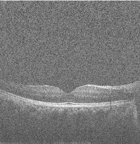
Application of Independent Component Analysis Techniques in Speckle Noise Reduction of Retinal OCT Images
Jul 28, 2015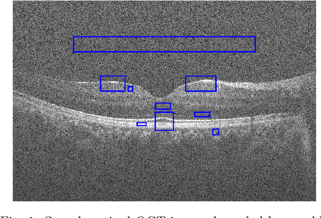
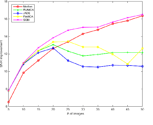
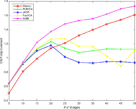
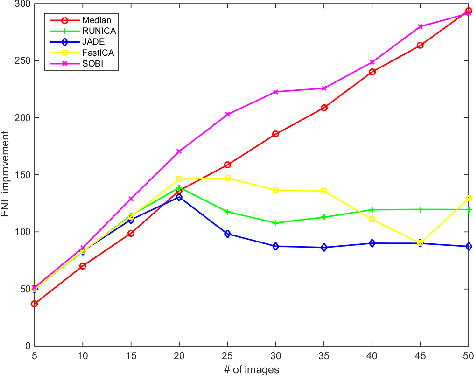
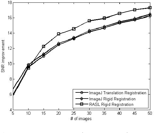
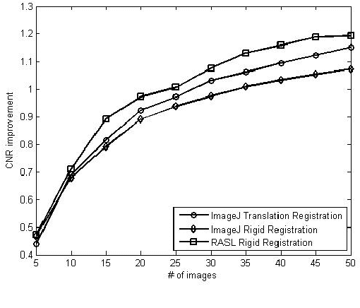
Optical Coherence Tomography (OCT) is an emerging technique in the field of biomedical imaging, with applications in ophthalmology, dermatology, coronary imaging etc. OCT images usually suffer from a granular pattern, called speckle noise, which restricts the process of interpretation. Therefore the need for speckle noise reduction techniques is of high importance. To the best of our knowledge, use of Independent Component Analysis (ICA) techniques has never been explored for speckle reduction of OCT images. Here, a comparative study of several ICA techniques (InfoMax, JADE, FastICA and SOBI) is provided for noise reduction of retinal OCT images. Having multiple B-scans of the same location, the eye movements are compensated using a rigid registration technique. Then, different ICA techniques are applied to the aggregated set of B-scans for extracting the noise-free image. Signal-to-Noise-Ratio (SNR), Contrast-to-Noise-Ratio (CNR) and Equivalent-Number-of-Looks (ENL), as well as analysis on the computational complexity of the methods, are considered as metrics for comparison. The results show that use of ICA can be beneficial, especially in case of having fewer number of B-scans.
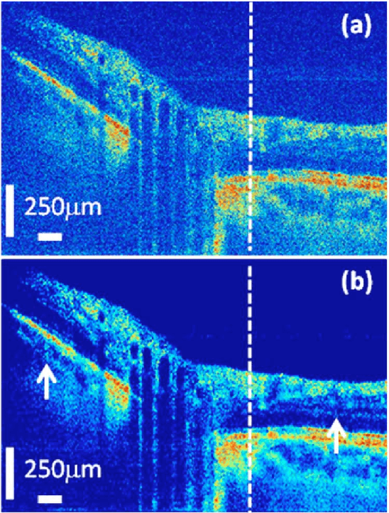
Sparse And Low Rank Decomposition Based Batch Image Alignment for Speckle Reduction of retinal OCT Images
Feb 09, 2015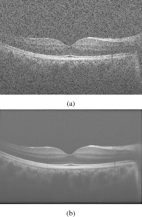
Optical Coherence Tomography (OCT) is an emerging technique in the field of biomedical imaging, with applications in ophthalmology, dermatology, coronary imaging etc. Due to the underlying physics, OCT images usually suffer from a granular pattern, called speckle noise, which restricts the process of interpretation. Here, a sparse and low rank decomposition based method is used for speckle reduction in retinal OCT images. This technique works on input data that consists of several B-scans of the same location. The next step is the batch alignment of the images using a sparse and low-rank decomposition based technique. Finally the denoised image is created by median filtering of the low-rank component of the processed data. Simultaneous decomposition and alignment of the images result in better performance in comparison to simple registration-based methods that are used in the literature for noise reduction of OCT images.
Structure Tensor Based Image Interpolation Method
Dec 26, 2014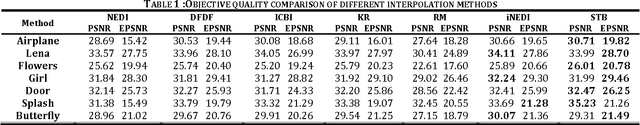
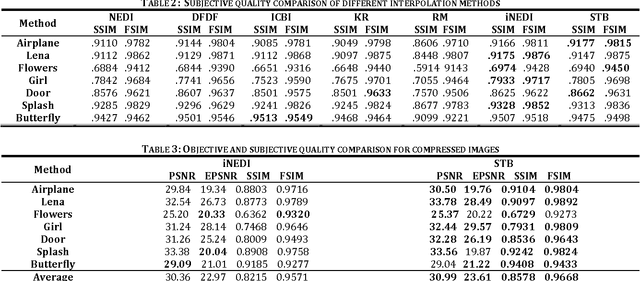
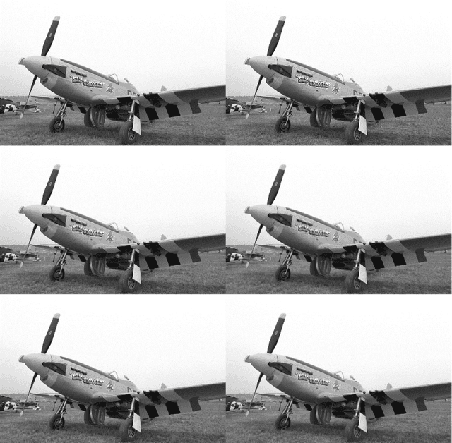

Feature preserving image interpolation is an active area in image processing field. In this paper a new direct edge directed image super-resolution algorithm based on structure tensors is proposed. Using an isotropic Gaussian filter, the structure tensor at each pixel of the input image is computed and the pixels are classified to three distinct classes; uniform region, corners and edges, according to the eigenvalues of the structure tensor. Due to application of the isotropic Gaussian filter, the classification is robust to noise presented in image. Based on the tangent eigenvector of the structure tensor, the edge direction is determined and used for interpolation along the edges. In comparison to some previous edge directed image interpolation methods, the proposed method achieves higher quality in both subjective and objective aspects. Also the proposed method outperforms previous methods in case of noisy and JPEG compressed images. Furthermore, without the need for optimization in the process, the algorithm can achieve higher speed.
State-of-the-Art in Retinal Optical Coherence Tomography Image Analysis
Nov 17, 2014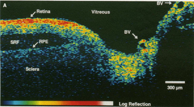
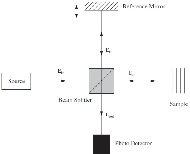
Optical Coherence Tomography (OCT) is one of the most emerging imaging modalities that has been used widely in the field of biomedical imaging. From its emergence in 1990's, plenty of hardware and software improvements have been made. Its applications range from ophthalmology to dermatology to coronary imaging etc. Here, the focus is on applications of OCT in ophthalmology and retinal imaging. OCT is able to non-invasively produce cross-sectional volume images of the tissues which are further used for analysis of the tissue structure and its properties. Due to the underlying physics, OCT images usually suffer from a granular pattern, called speckle noise, which restricts the process of interpretation, hence requiring specialized noise reduction techniques to remove the noise while preserving image details. Also, given the fact that OCT images are in the $\mu m$ -level, further analysis in needed to distinguish between the different structures in the imaged volume. Therefore the use of different segmentation techniques are of high importance. The movement of the tissue under imaging or the progression of disease in the tissue also imposes further implications both on the quality and the proper interpretation of the acquired images. Thus, use of image registration techniques can be very helpful. In this work, an overview of such image analysis techniques will be given.
Fast Mesh-Based Medical Image Registration
Nov 08, 2014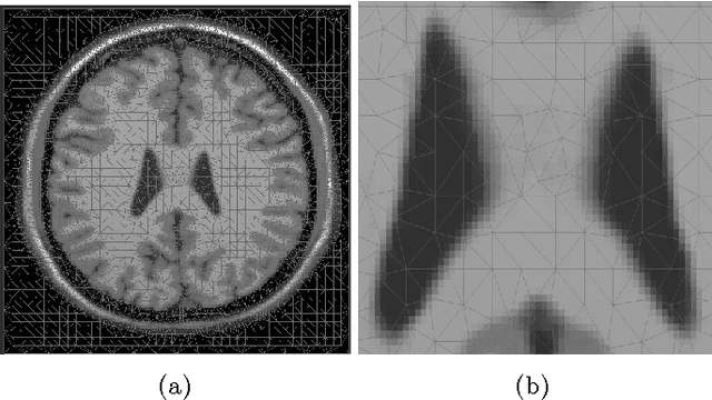
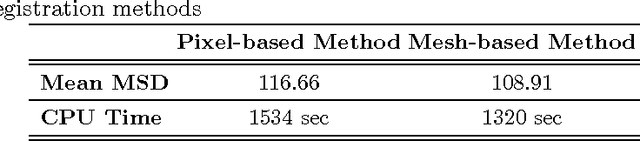
In this paper a fast triangular mesh based registration method is proposed. Having Template and Reference images as inputs, the template image is triangulated using a content adaptive mesh generation algorithm. Considering the pixel values at mesh nodes, interpolated using spline interpolation method for both of the images, the energy functional needed for image registration is minimized. The minimization process was achieved using a mesh based discretization of the distance measure and regularization term which resulted in a sparse system of linear equations, which due to the smaller size in comparison to the pixel-wise registration method, can be solved directly. Mean Squared Difference (MSD) is used as a metric for evaluating the results. Using the mesh based technique, higher speed was achieved compared to pixel-based curvature registration technique with fast DCT solver. The implementation was done in MATLAB without any specific optimization. Higher speeds can be achieved using C/C++ implementations.
An Optimization Method For Slice Interpolation Of Medical Images
Mar 27, 2014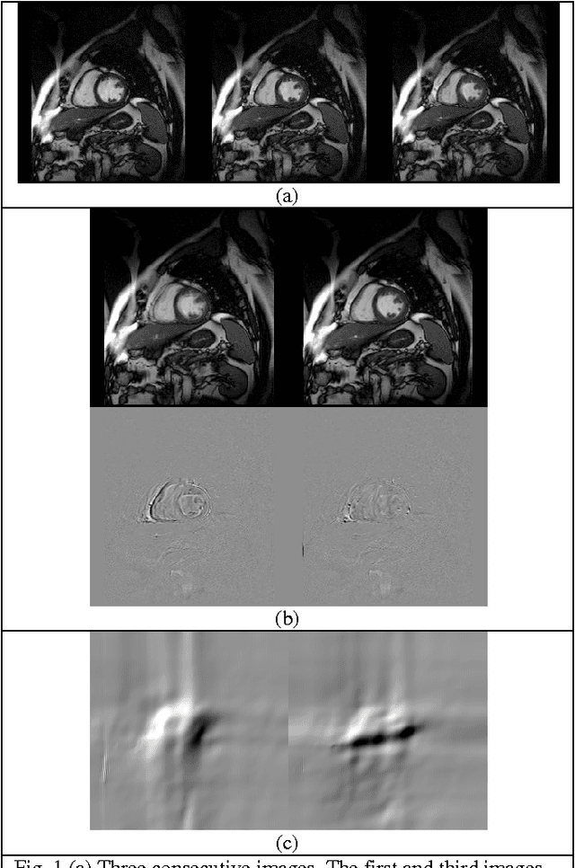
Slice interpolation is a fast growing field in medical image processing. Intensity-based interpolation and object-based interpolation are two major groups of methods in the literature. In this paper, we describe an object-oriented, optimization method based on a modified version of curvature-based image registration, in which a displacement field is computed for the missing slice between two known slices and used to interpolate the intensities of the missing slice. The proposed approach is evaluated quantitatively by using the Mean Squared Difference (MSD) as a metric. The produced results also show visual improvement in preserving sharp edges in images.