 Add to Chrome
 Add to Firefox
 Add to Edge
SAGE: Semantic Explainability of Attention-Based Survival Models in Computational Pathology
Aug 03, 2026
Attention-based multiple instance learning (ABMIL) is the predominant approach for slide-level prediction in computational pathology, yet its attention maps provide only local explanations: they indicate where a model focuses but not which histological features drive its predictions or how the model behaves across a patient cohort. We present Semantic Attention Global Explanations (SAGE), a post-hoc framework that extracts global, language-grounded explanations from a frozen ABMIL model. Using a pathology vision-language model, SAGE scores image patches against a dictionary of 25 histological concepts, aggregates these scores according to the model's learned attention, and quantifies how each concept relates to prediction risk across a cohort. Applied to survival prediction using seven TCGA cancer cohorts and three foundation models, SAGE recovered established prognostic features, such as the adverse association of necrosis, while revealing cancer-specific biology, including a favorable angiogenic signature in renal cell carcinoma consistent with known molecular subtypes. Ablation studies demonstrated that these associations depend on the model's learned attention rather than concept prevalence alone, and that the concept dictionary captures much of the prognostic information encoded by the foundation model features. Through semantically-grounded explanations, SAGE provides a scalable, model-agnostic framework for understanding what ABMIL survival models learn, enabling pathologists to interpret model behavior at the cohort level and offering the potential for biomarker identification.

Anatomy Contextualized Adaption of CT Foundation Models
Jul 29, 2026CT vision-language foundation models have demonstrated promising performance across downstream tasks, but are typically trained with whole-volume representations that dilute fine-grained anatomical signals. Fine-grained vision-language pre-training addresses this by aligning anatomy-level visual features with anatomy-specific text, but in doing so discards the global context that whole-volume models provide. Furthermore, existing fine-grained approaches train from scratch, making them computationally expensive. We introduce Anatomy Contextualized Adaptation (ACA), a lightweight framework that adapts frozen CT foundation model representations for anatomy-level vision-language alignment while enhancing global contextualization. ACA uses TotalSegmentator to decompose CT volumes into anatomy-level embeddings, which are refined via a transformer that captures cross-anatomy relationships, and aligned to both per-anatomy and scan-level text extracted from radiology reports. Evaluated on Merlin and CT-RATE, ACA consistently outperforms both the frozen foundation model baselines and existing fine-grained methods in zero-shot finding classification, while requiring less than one hour of training once embeddings are cached. The attention weights learned by ACA's inter-anatomy transformer additionally indicate plausible cross-anatomy context routing. Altogether, these results support ACA as a lightweight approach for adapting CT foundation models to anatomically grounded vision-language alignment while preserving and enhancing global anatomical context.
How Seemingly Inconsequential Design Choices Dictate Performance of LLMs in Pathology
Jun 10, 2026General-purpose large language models (LLMs) are routinely used as baselines when evaluating specialized pathology models on whole-slide images (WSIs). Because WSIs exceed contemporary model context limits, LLM baselines routinely use small, high-magnification patches processed independently via majority voting, without systematic evaluation of seemingly inconsequential design choices such as patch size, patch count, and magnification. Generalist LLMs have consistently underperformed specialized systems, reinforcing the perception that domain-specific training or architectural adaptation is necessary for pathology tasks involving WSIs. Here, we conduct a systematic factorial analysis of four input design factors: inference mode, patch size, magnification, and patch count. We demonstrate that prior studies have overstated the gap between specialized models and general-purpose LLMs by choosing non-optimized input configurations. On the MultiPathQA benchmark, switching to a single balanced configuration (large patches at lower magnification, processed jointly) raises GPT-5 from 15.1% to 39.5% on cancer-type classification (TCGA) and from 38.1% to 62.9% on organ classification (GTEx). Per-task optimization yields further gains up to 43.9% (TCGA) and 71.6% (GTEx). The same configuration generalizes to two other models and to a fully held-out CPTAC cohort, where it improves Gemini 3 Flash by 23.4 percentage points without any task-specific tuning.
Evaluating the Impact of Medical Image Reconstruction on Downstream AI Fairness and Performance
Apr 13, 2026AI-based image reconstruction models are increasingly deployed in clinical workflows to improve image quality from noisy data, such as low-dose X-rays or accelerated MRI scans. However, these models are typically evaluated using pixel-level metrics like PSNR, leaving their impact on downstream diagnostic performance and fairness unclear. We introduce a scalable evaluation framework that applies reconstruction and diagnostic AI models in tandem, which we apply to two tasks (classification, segmentation), three reconstruction approaches (U-Net, GAN, diffusion), and two data types (X-ray, MRI) to assess the potential downstream implications of reconstruction. We find that conventional reconstruction metrics poorly track task performance, where diagnostic accuracy remains largely stable even as reconstruction PSNR declines with increasing image noise. Fairness metrics exhibit greater variability, with reconstruction sometimes amplifying demographic biases, particularly regarding patient sex. However, the overall magnitude of this additional bias is modest compared to the inherent biases already present in diagnostic models. To explore potential bias mitigation, we adapt two strategies from classification literature to the reconstruction setting, but observe limited efficacy. Overall, our findings emphasize the importance of holistic performance and fairness assessments throughout the entire medical imaging workflow, especially as generative reconstruction models are increasingly deployed.
Do Pathology Foundation Models Encode Disease Progression? A Pseudotime Analysis of Visual Representations
Jan 29, 2026Vision foundation models trained on discretely sampled images achieve strong performance on classification benchmarks, yet whether their representations encode the continuous processes underlying their training data remains unclear. This question is especially pertinent in computational pathology, where we posit that models whose latent representations implicitly capture continuous disease progression may better reflect underlying biology, support more robust generalization, and enable quantitative analyses of features associated with disease transitions. Using diffusion pseudotime, a method developed to infer developmental trajectories from single-cell transcriptomics, we probe whether foundation models organize disease states along coherent progression directions in representation space. Across four cancer progressions and six models, we find that all pathology-specific models recover trajectory orderings significantly exceeding null baselines, with vision-only models achieving the highest fidelities $(τ> 0.78$ on CRC-Serrated). Model rankings by trajectory fidelity on reference diseases strongly predict few-shot classification performance on held-out diseases ($ρ= 0.92$), and exploratory analysis shows cell-type composition varies smoothly along inferred trajectories in patterns consistent with known stromal remodeling. Together, these results demonstrate that vision foundation models can implicitly learn to represent continuous processes from independent static observations, and that trajectory fidelity provides a complementary measure of representation quality beyond downstream performance. While demonstrated in pathology, this framework could be applied to other domains where continuous processes are observed through static snapshots.
Comparing Computational Pathology Foundation Models using Representational Similarity Analysis
Sep 18, 2025Foundation models are increasingly developed in computational pathology (CPath) given their promise in facilitating many downstream tasks. While recent studies have evaluated task performance across models, less is known about the structure and variability of their learned representations. Here, we systematically analyze the representational spaces of six CPath foundation models using techniques popularized in computational neuroscience. The models analyzed span vision-language contrastive learning (CONCH, PLIP, KEEP) and self-distillation (UNI (v2), Virchow (v2), Prov-GigaPath) approaches. Through representational similarity analysis using H&E image patches from TCGA, we find that UNI2 and Virchow2 have the most distinct representational structures, whereas Prov-Gigapath has the highest average similarity across models. Having the same training paradigm (vision-only vs. vision-language) did not guarantee higher representational similarity. The representations of all models showed a high slide-dependence, but relatively low disease-dependence. Stain normalization decreased slide-dependence for all models by a range of 5.5% (CONCH) to 20.5% (PLIP). In terms of intrinsic dimensionality, vision-language models demonstrated relatively compact representations, compared to the more distributed representations of vision-only models. These findings highlight opportunities to improve robustness to slide-specific features, inform model ensembling strategies, and provide insights into how training paradigms shape model representations. Our framework is extendable across medical imaging domains, where probing the internal representations of foundation models can help ensure effective development and deployment.
Simulating Clinical AI Assistance using Multimodal LLMs: A Case Study in Diabetic Retinopathy
Sep 16, 2025Diabetic retinopathy (DR) is a leading cause of blindness worldwide, and AI systems can expand access to fundus photography screening. Current FDA-cleared systems primarily provide binary referral outputs, where this minimal output may limit clinical trust and utility. Yet, determining the most effective output format to enhance clinician-AI performance is an empirical challenge that is difficult to assess at scale. We evaluated multimodal large language models (MLLMs) for DR detection and their ability to simulate clinical AI assistance across different output types. Two models were tested on IDRiD and Messidor-2: GPT-4o, a general-purpose MLLM, and MedGemma, an open-source medical model. Experiments included: (1) baseline evaluation, (2) simulated AI assistance with synthetic predictions, and (3) actual AI-to-AI collaboration where GPT-4o incorporated MedGemma outputs. MedGemma outperformed GPT-4o at baseline, achieving higher sensitivity and AUROC, while GPT-4o showed near-perfect specificity but low sensitivity. Both models adjusted predictions based on simulated AI inputs, but GPT-4o's performance collapsed with incorrect ones, whereas MedGemma remained more stable. In actual collaboration, GPT-4o achieved strong results when guided by MedGemma's descriptive outputs, even without direct image access (AUROC up to 0.96). These findings suggest MLLMs may improve DR screening pipelines and serve as scalable simulators for studying clinical AI assistance across varying output configurations. Open, lightweight models such as MedGemma may be especially valuable in low-resource settings, while descriptive outputs could enhance explainability and clinician trust in clinical workflows.
AdvDINO: Domain-Adversarial Self-Supervised Representation Learning for Spatial Proteomics
Aug 07, 2025Self-supervised learning (SSL) has emerged as a powerful approach for learning visual representations without manual annotations. However, the robustness of standard SSL methods to domain shift -- systematic differences across data sources -- remains uncertain, posing an especially critical challenge in biomedical imaging where batch effects can obscure true biological signals. We present AdvDINO, a domain-adversarial self-supervised learning framework that integrates a gradient reversal layer into the DINOv2 architecture to promote domain-invariant feature learning. Applied to a real-world cohort of six-channel multiplex immunofluorescence (mIF) whole slide images from non-small cell lung cancer patients, AdvDINO mitigates slide-specific biases to learn more robust and biologically meaningful representations than non-adversarial baselines. Across $>5.46$ million mIF image tiles, the model uncovers phenotype clusters with distinct proteomic profiles and prognostic significance, and improves survival prediction in attention-based multiple instance learning. While demonstrated on mIF data, AdvDINO is broadly applicable to other imaging domains -- including radiology, remote sensing, and autonomous driving -- where domain shift and limited annotated data hinder model generalization and interpretability.
Fluoroformer: Scaling multiple instance learning to multiplexed images via attention-based channel fusion
Nov 13, 2024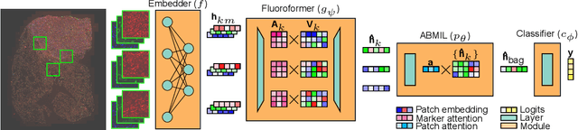
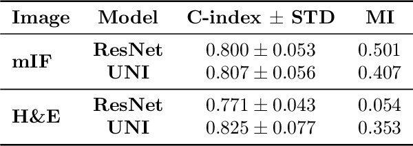
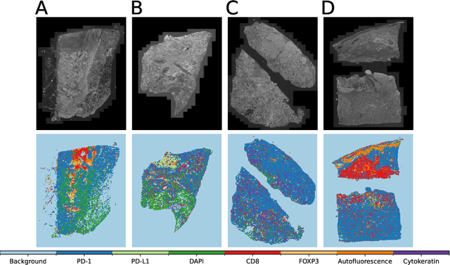

Though multiple instance learning (MIL) has been a foundational strategy in computational pathology for processing whole slide images (WSIs), current approaches are designed for traditional hematoxylin and eosin (H&E) slides rather than emerging multiplexed technologies. Here, we present an MIL strategy, the Fluoroformer module, that is specifically tailored to multiplexed WSIs by leveraging scaled dot-product attention (SDPA) to interpretably fuse information across disparate channels. On a cohort of 434 non-small cell lung cancer (NSCLC) samples, we show that the Fluoroformer both obtains strong prognostic performance and recapitulates immuno-oncological hallmarks of NSCLC. Our technique thereby provides a path for adapting state-of-the-art AI techniques to emerging spatial biology assays.
Representing visual classification as a linear combination of words
Nov 18, 2023
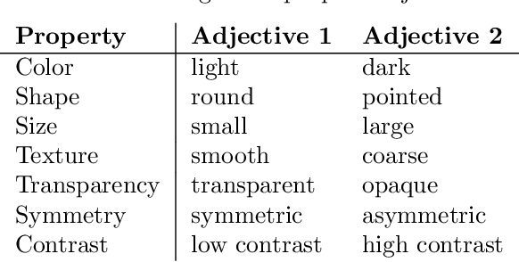
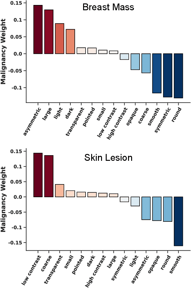
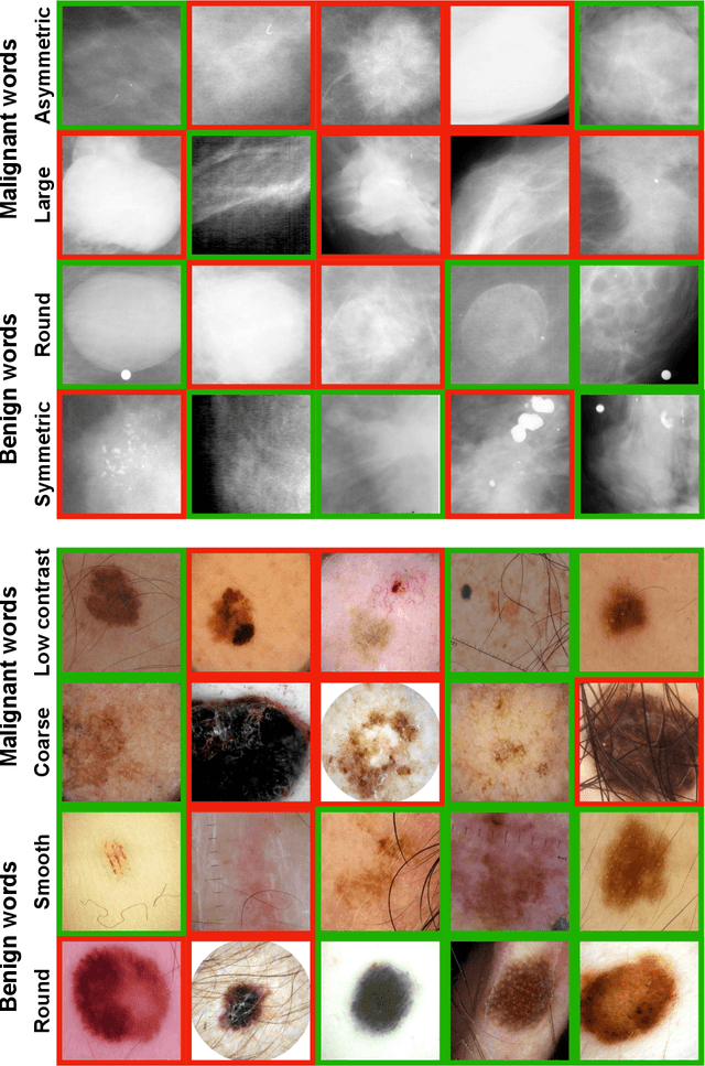
Explainability is a longstanding challenge in deep learning, especially in high-stakes domains like healthcare. Common explainability methods highlight image regions that drive an AI model's decision. Humans, however, heavily rely on language to convey explanations of not only "where" but "what". Additionally, most explainability approaches focus on explaining individual AI predictions, rather than describing the features used by an AI model in general. The latter would be especially useful for model and dataset auditing, and potentially even knowledge generation as AI is increasingly being used in novel tasks. Here, we present an explainability strategy that uses a vision-language model to identify language-based descriptors of a visual classification task. By leveraging a pre-trained joint embedding space between images and text, our approach estimates a new classification task as a linear combination of words, resulting in a weight for each word that indicates its alignment with the vision-based classifier. We assess our approach using two medical imaging classification tasks, where we find that the resulting descriptors largely align with clinical knowledge despite a lack of domain-specific language training. However, our approach also identifies the potential for 'shortcut connections' in the public datasets used. Towards a functional measure of explainability, we perform a pilot reader study where we find that the AI-identified words can enable non-expert humans to perform a specialized medical task at a non-trivial level. Altogether, our results emphasize the potential of using multimodal foundational models to deliver intuitive, language-based explanations of visual tasks.