 Add to Chrome
 Add to Firefox
 Add to Edge
ConvFormer3D-TAP: Phase/Uncertainty-Aware Front-End Fusion for Cine CMR View Classification Pipelines
Apr 13, 2026
Reliable recognition of standard cine cardiac MRI views is essential because each view determines which cardiac anatomy is visualized and which quantitative analyses can be performed. Incorrect view identification, whether by a human reader or an automated deep learning system, can propagate errors into segmentation, volumetric assessment, strain analysis, and valve evaluation. However, accurate view classification remains challenging under routine clinical variability in scanner vendor, acquisition protocol, motion artifacts, and plane prescription. We present ConvFormer3D-TAP, a cine-specific spatiotemporal architecture that integrates 3D convolutional tokenization with multiscale self-attention. The model is trained using masked spatiotemporal reconstruction and uncertainty-weighted multi-clip fusion to enhance robustness across cardiac phases and ambiguous temporal segments. The design captures complementary cues: local anatomical structure through convolutional priors and long-range cardiac-cycle dynamics through hierarchical attention. On a cohort of 150,974 clinically acquired cine sequences spanning six standard cine cardiac MRI views, ConvFormer3D-TAP achieved 96% validation accuracy with per-class F1-scores >= 0.94 and strong calibration (ECE = 0.025; Brier = 0.040). Error analysis shows that residual confusions are concentrated in anatomically adjacent long-axis and LVOT/AV view pairs, consistent with intrinsic prescription overlap. These results support ConvFormer3D-TAP as a scalable front-end for view routing, filtering and quality control in end-to-end cMRI workflows.

Medical Image Deidentification, Cleaning and Compression Using Pylogik
May 03, 2023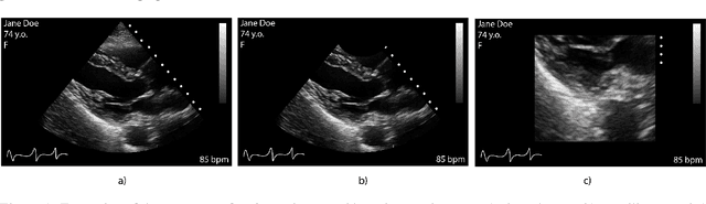


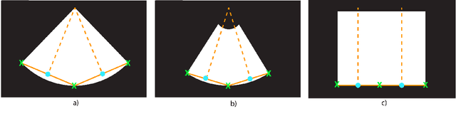
Leveraging medical record information in the era of big data and machine learning comes with the caveat that data must be cleaned and deidentified. Facilitating data sharing and harmonization for multi-center collaborations are particularly difficult when protected health information (PHI) is contained or embedded in image meta-data. We propose a novel library in the Python framework, called PyLogik, to help alleviate this issue for ultrasound images, which are particularly challenging because of the frequent inclusion of PHI directly on the images. PyLogik processes the image volumes through a series of text detection/extraction, filtering, thresholding, morphological and contour comparisons. This methodology deidentifies the images, reduces file sizes, and prepares image volumes for applications in deep learning and data sharing. To evaluate its effectiveness in the identification of regions of interest (ROI), a random sample of 50 cardiac ultrasounds (echocardiograms) were processed through PyLogik, and the outputs were compared with the manual segmentations by an expert user. The Dice coefficient of the two approaches achieved an average value of 0.976. Next, an investigation was conducted to ascertain the degree of information compression achieved using the algorithm. Resultant data was found to be on average approximately 72% smaller after processing by PyLogik. Our results suggest that PyLogik is a viable methodology for ultrasound data cleaning and deidentification, determining ROI, and file compression which will facilitate efficient storage, use, and dissemination of ultrasound data.