 Add to Chrome
 Add to Firefox
 Add to Edge
Detecting Shortcuts in Medical Images -- A Case Study in Chest X-rays
Nov 09, 2022The availability of large public datasets and the increased amount of computing power have shifted the interest of the medical community to high-performance algorithms. However, little attention is paid to the quality of the data and their annotations. High performance on benchmark datasets may be reported without considering possible shortcuts or artifacts in the data, besides, models are not tested on subpopulation groups. With this work, we aim to raise awareness about shortcuts problems. We validate previous findings, and present a case study on chest X-rays using two publicly available datasets. We share annotations for a subset of pneumothorax images with drains. We conclude with general recommendations for medical image classification.
Detection of Furigana Text in Images
Jul 08, 2022
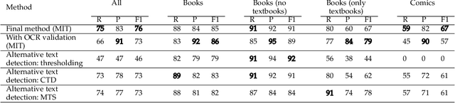

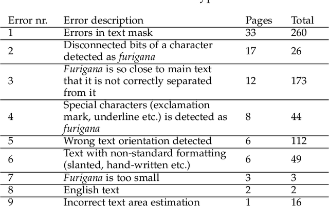
Furigana are pronunciation notes used in Japanese writing. Being able to detect these can help improve optical character recognition (OCR) performance or make more accurate digital copies of Japanese written media by correctly displaying furigana. This project focuses on detecting furigana in Japanese books and comics. While there has been research into the detection of Japanese text in general, there are currently no proposed methods for detecting furigana. We construct a new dataset containing Japanese written media and annotations of furigana. We propose an evaluation metric for such data which is similar to the evaluation protocols used in object detection except that it allows groups of objects to be labeled by one annotation. We propose a method for detection of furigana that is based on mathematical morphology and connected component analysis. We evaluate the detections of the dataset and compare different methods for text extraction. We also evaluate different types of images such as books and comics individually and discuss the challenges of each type of image. The proposed method reaches an F1-score of 76\% on the dataset. The method performs well on regular books, but less so on comics, and books of irregular format. Finally, we show that the proposed method can improve the performance of OCR by 5\% on the manga109 dataset. Source code is available via \texttt{\url{https://github.com/nikolajkb/FuriganaDetection}}
Metrics reloaded: Pitfalls and recommendations for image analysis validation
Jun 03, 2022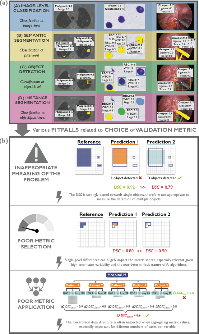
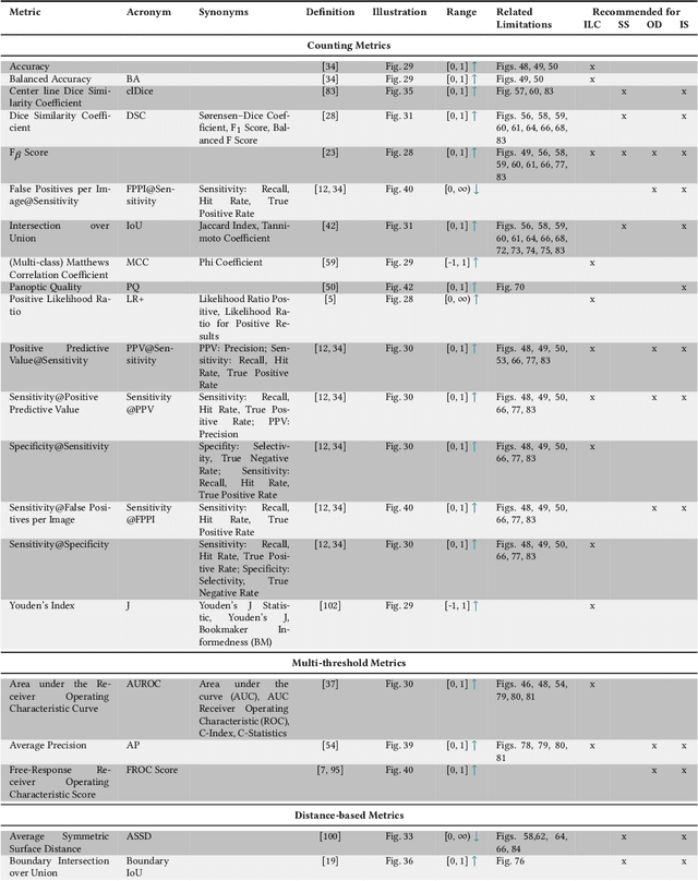
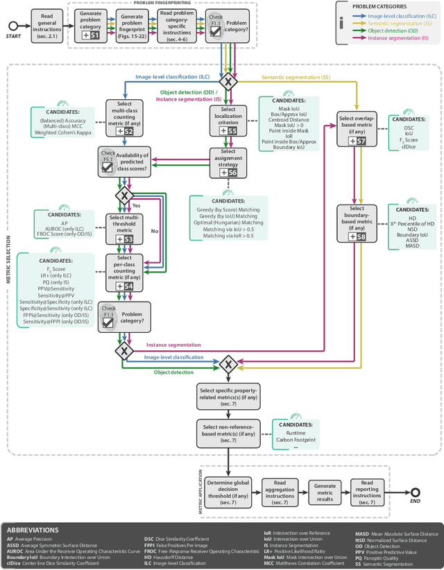
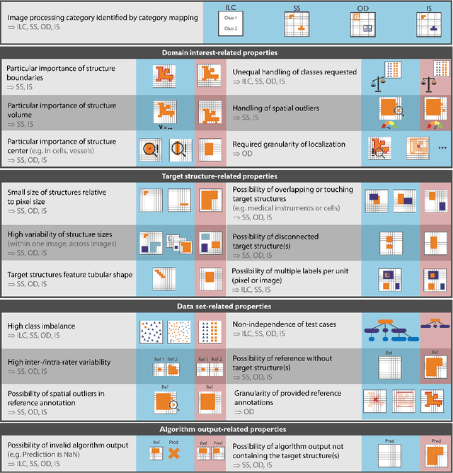
The field of automatic biomedical image analysis crucially depends on robust and meaningful performance metrics for algorithm validation. Current metric usage, however, is often ill-informed and does not reflect the underlying domain interest. Here, we present a comprehensive framework that guides researchers towards choosing performance metrics in a problem-aware manner. Specifically, we focus on biomedical image analysis problems that can be interpreted as a classification task at image, object or pixel level. The framework first compiles domain interest-, target structure-, data set- and algorithm output-related properties of a given problem into a problem fingerprint, while also mapping it to the appropriate problem category, namely image-level classification, semantic segmentation, instance segmentation, or object detection. It then guides users through the process of selecting and applying a set of appropriate validation metrics while making them aware of potential pitfalls related to individual choices. In this paper, we describe the current status of the Metrics Reloaded recommendation framework, with the goal of obtaining constructive feedback from the image analysis community. The current version has been developed within an international consortium of more than 60 image analysis experts and will be made openly available as a user-friendly toolkit after community-driven optimization.
Predicting Bearings' Degradation Stages for Predictive Maintenance in the Pharmaceutical Industry
Mar 07, 2022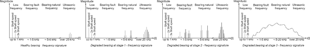
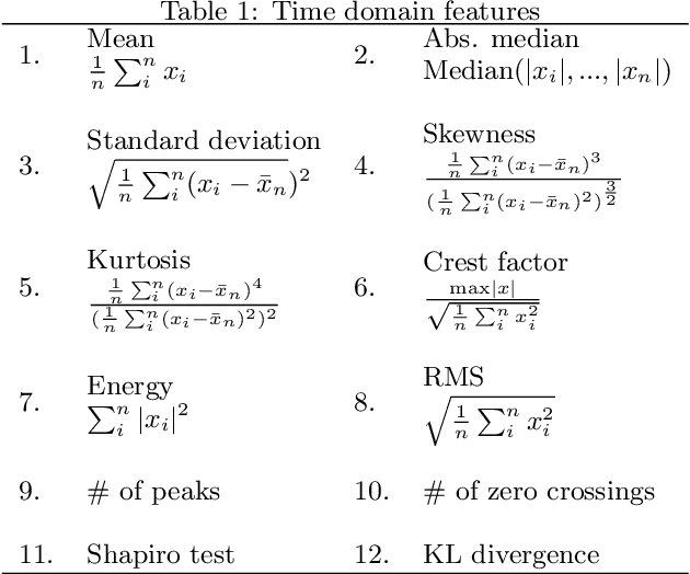
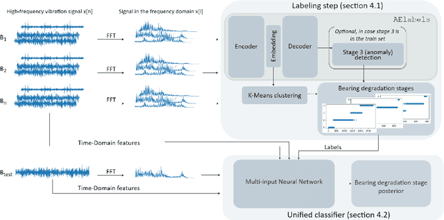
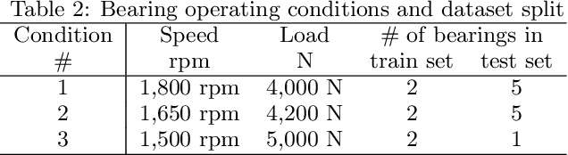
In the pharmaceutical industry, the maintenance of production machines must be audited by the regulator. In this context, the problem of predictive maintenance is not when to maintain a machine, but what parts to maintain at a given point in time. The focus shifts from the entire machine to its component parts and prediction becomes a classification problem. In this paper, we focus on rolling-elements bearings and we propose a framework for predicting their degradation stages automatically. Our main contribution is a k-means bearing lifetime segmentation method based on high-frequency bearing vibration signal embedded in a latent low-dimensional subspace using an AutoEncoder. Given high-frequency vibration data, our framework generates a labeled dataset that is used to train a supervised model for bearing degradation stage detection. Our experimental results, based on the FEMTO Bearing dataset, show that our framework is scalable and that it provides reliable and actionable predictions for a range of different bearings.
Effect of Prior-based Losses on Segmentation Performance: A Benchmark
Jan 12, 2022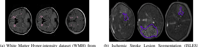
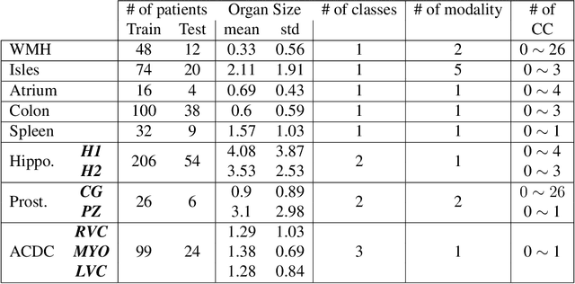
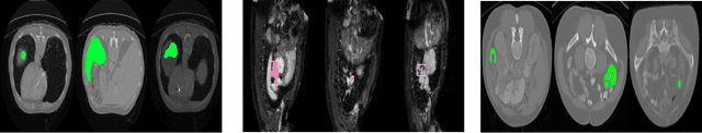
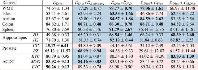
Today, deep convolutional neural networks (CNNs) have demonstrated state-of-the-art performance for medical image segmentation, on various imaging modalities and tasks. Despite early success, segmentation networks may still generate anatomically aberrant segmentations, with holes or inaccuracies near the object boundaries. To enforce anatomical plausibility, recent research studies have focused on incorporating prior knowledge such as object shape or boundary, as constraints in the loss function. Prior integrated could be low-level referring to reformulated representations extracted from the ground-truth segmentations, or high-level representing external medical information such as the organ's shape or size. Over the past few years, prior-based losses exhibited a rising interest in the research field since they allow integration of expert knowledge while still being architecture-agnostic. However, given the diversity of prior-based losses on different medical imaging challenges and tasks, it has become hard to identify what loss works best for which dataset. In this paper, we establish a benchmark of recent prior-based losses for medical image segmentation. The main objective is to provide intuition onto which losses to choose given a particular task or dataset. To this end, four low-level and high-level prior-based losses are selected. The considered losses are validated on 8 different datasets from a variety of medical image segmentation challenges including the Decathlon, the ISLES and the WMH challenge. Results show that whereas low-level prior-based losses can guarantee an increase in performance over the Dice loss baseline regardless of the dataset characteristics, high-level prior-based losses can increase anatomical plausibility as per data characteristics.
ENHANCE : A case study for skin lesion classification
Jul 27, 2021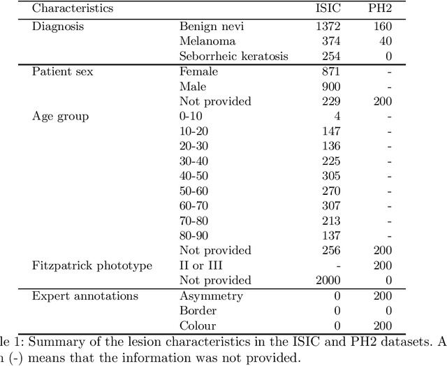
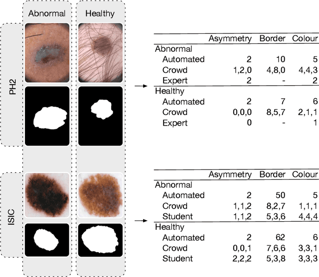
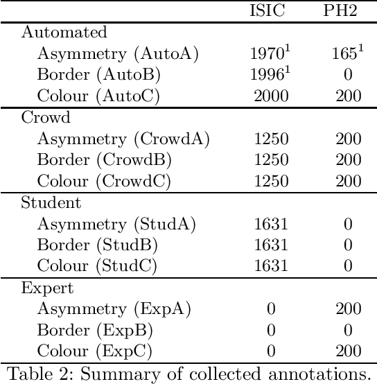
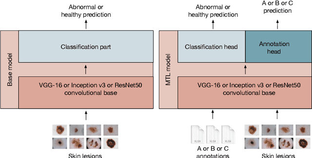
We present ENHANCE, an open dataset with multiple annotations to complement the existing ISIC and PH2 skin lesion classification datasets. This dataset contains annotations of visual ABC (asymmetry, border, colour) features from non-expert annotation sources: undergraduate students, crowd workers from Amazon MTurk and classic image processing algorithms. In this paper we first analyse the correlations between the annotations and the diagnostic label of the lesion, as well as study the agreement between different annotation sources. Overall we find weak correlations of non-expert annotations with the diagnostic label, and low agreement between different annotation sources. We then study multi-task learning (MTL) with the annotations as additional labels, and show that non-expert annotations can improve (ensembles of) state-of-the-art convolutional neural networks via MTL. We hope that our dataset can be used in further research into multiple annotations and/or MTL. All data and models are available on Github: https://github.com/raumannsr/ENHANCE.
Cats, not CAT scans: a study of dataset similarity in transfer learning for 2D medical image classification
Jul 13, 2021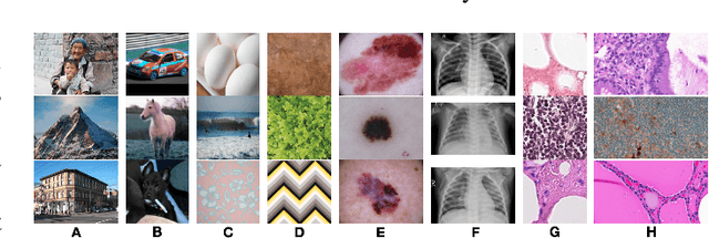
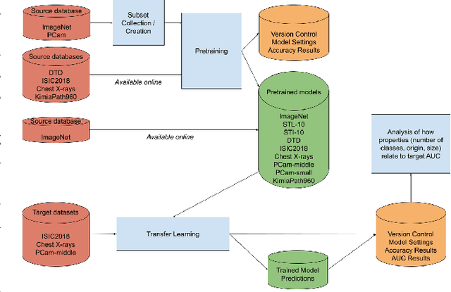
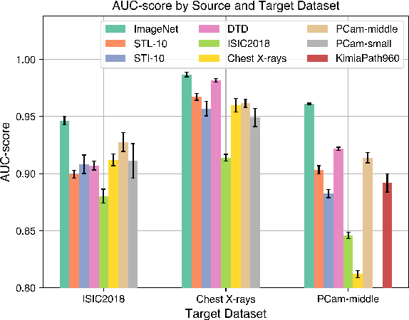
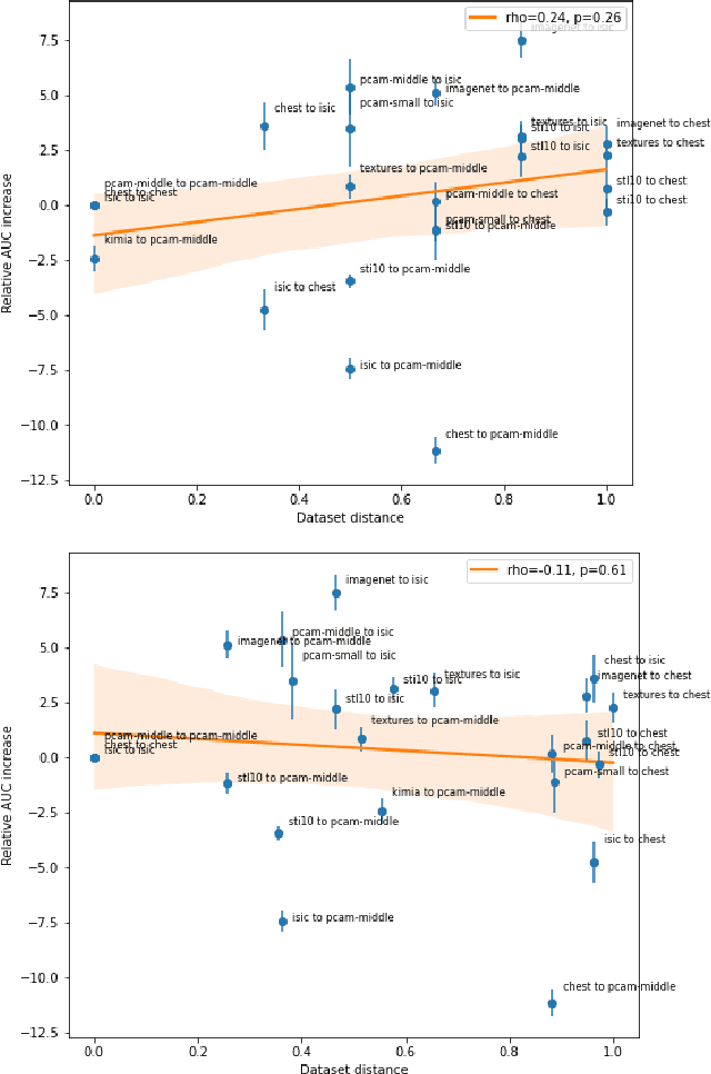
Transfer learning is a commonly used strategy for medical image classification, especially via pretraining on source data and fine-tuning on target data. There is currently no consensus on how to choose appropriate source data, and in the literature we can find both evidence of favoring large natural image datasets such as ImageNet, and evidence of favoring more specialized medical datasets. In this paper we perform a systematic study with nine source datasets with natural or medical images, and three target medical datasets, all with 2D images. We find that ImageNet is the source leading to the highest performances, but also that larger datasets are not necessarily better. We also study different definitions of data similarity. We show that common intuitions about similarity may be inaccurate, and therefore not sufficient to predict an appropriate source a priori. Finally, we discuss several steps needed for further research in this field, especially with regard to other types (for example 3D) medical images. Our experiments and pretrained models are available via \url{https://www.github.com/vcheplygina/cats-scans}
Common Limitations of Image Processing Metrics: A Picture Story
Apr 13, 2021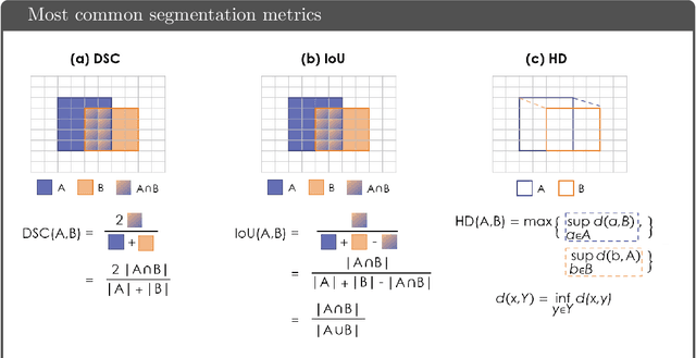
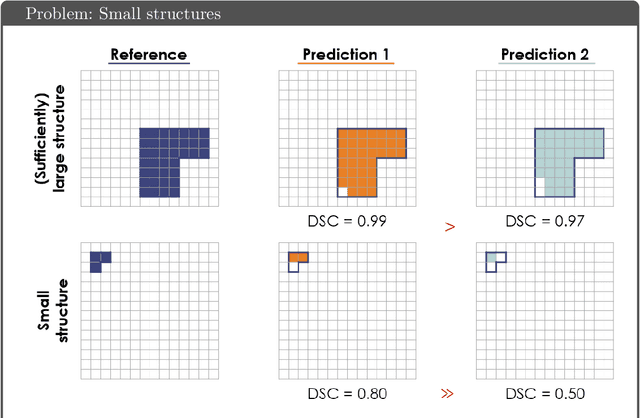
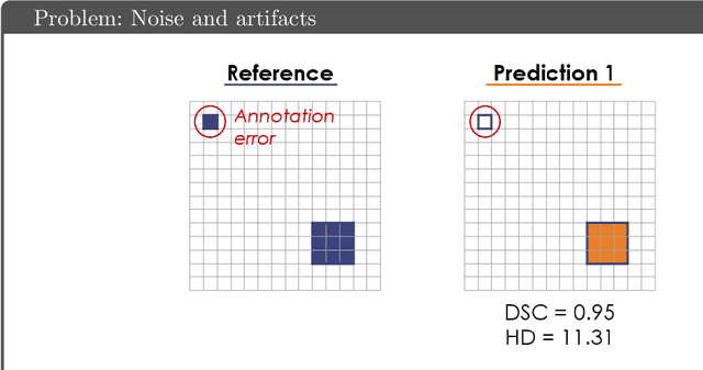
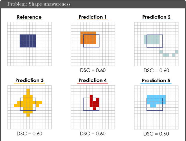
While the importance of automatic image analysis is increasing at an enormous pace, recent meta-research revealed major flaws with respect to algorithm validation. Specifically, performance metrics are key for objective, transparent and comparative performance assessment, but relatively little attention has been given to the practical pitfalls when using specific metrics for a given image analysis task. A common mission of several international initiatives is therefore to provide researchers with guidelines and tools to choose the performance metrics in a problem-aware manner. This dynamically updated document has the purpose to illustrate important limitations of performance metrics commonly applied in the field of image analysis. The current version is based on a Delphi process on metrics conducted by an international consortium of image analysis experts.
How I failed machine learning in medical imaging -- shortcomings and recommendations
Mar 18, 2021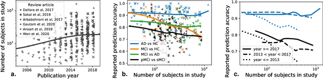
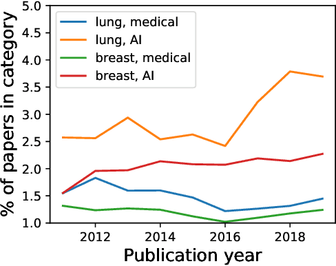
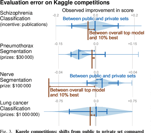
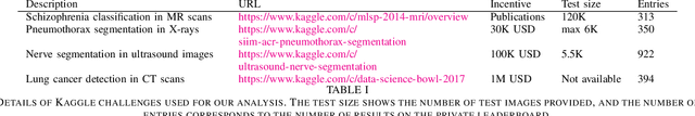
Medical imaging is an important research field with many opportunities for improving patients' health. However, there are a number of challenges that are slowing down the progress of the field as a whole, such optimizing for publication. In this paper we reviewed several problems related to choosing datasets, methods, evaluation metrics, and publication strategies. With a review of literature and our own analysis, we show that at every step, potential biases can creep in. On a positive note, we also see that initiatives to counteract these problems are already being started. Finally we provide a broad range of recommendations on how to further these address problems in the future. For reproducibility, data and code for our analyses are available on \url{https://github.com/GaelVaroquaux/ml_med_imaging_failures}
Using uncertainty estimation to reduce false positives in liver lesion detection
Jan 26, 2021
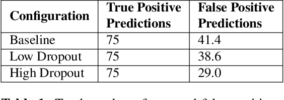
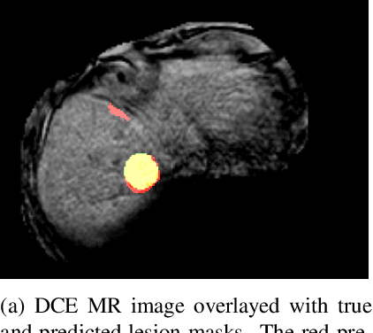
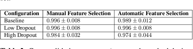
Despite the successes of deep learning techniques at detecting objects in medical images, false positive detections occur which may hinder an accurate diagnosis. We propose a technique to reduce false positive detections made by a neural network using an SVM classifier trained with features derived from the uncertainty map of the neural network prediction. We demonstrate the effectiveness of this method for the detection of liver lesions on a dataset of abdominal MR images. We find that the use of a dropout rate of 0.5 produces the least number of false positives in the neural network predictions and the trained classifier filters out approximately 90% of these false positives detections in the test-set.