 Add to Chrome
 Add to Firefox
 Add to Edge
Predicting ulcer in H&E images of inflammatory bowel disease using domain-knowledge-driven graph neural network
Apr 13, 2025Inflammatory bowel disease (IBD) involves chronic inflammation of the digestive tract, with treatment options often burdened by adverse effects. Identifying biomarkers for personalized treatment is crucial. While immune cells play a key role in IBD, accurately identifying ulcer regions in whole slide images (WSIs) is essential for characterizing these cells and exploring potential therapeutics. Multiple instance learning (MIL) approaches have advanced WSI analysis but they lack spatial context awareness. In this work, we propose a weakly-supervised model called DomainGCN that employs a graph convolution neural network (GCN) and incorporates domain-specific knowledge of ulcer features, specifically, the presence of epithelium, lymphocytes, and debris for WSI-level ulcer prediction in IBD. We demonstrate that DomainGCN outperforms various state-of-the-art (SOTA) MIL methods and show the added value of domain knowledge.
Restoration of marker occluded hematoxylin and eosin stained whole slide histology images using generative adversarial networks
Oct 14, 2019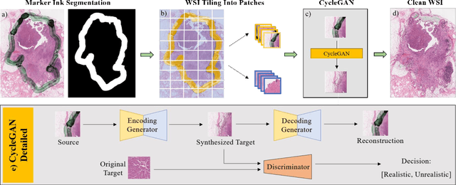
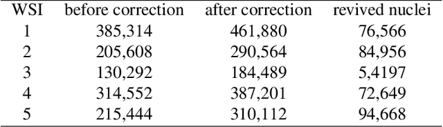
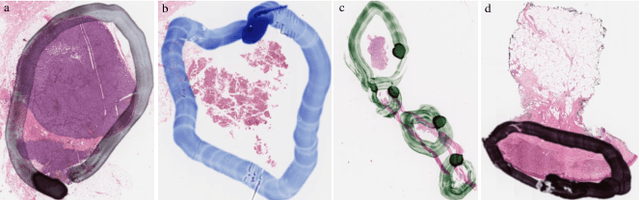
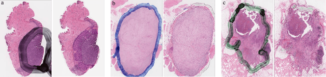
It is common for pathologists to annotate specific regions of the tissue, such as tumor, directly on the glass slide with markers. Although this practice was helpful prior to the advent of histology whole slide digitization, it often occludes important details which are increasingly relevant to immuno-oncology due to recent advancements in digital pathology imaging techniques. The current work uses a generative adversarial network with cycle loss to remove these annotations while still maintaining the underlying structure of the tissue by solving an image-to-image translation problem. We train our network on up to 300 whole slide images with marker inks and show that 70% of the corrected image patches are indistinguishable from originally uncontaminated image tissue to a human expert. This portion increases 97% when we replace the human expert with a deep residual network. We demonstrated the fidelity of the method to the original image by calculating the correlation between image gradient magnitudes. We observed a revival of up to 94,000 nuclei per slide in our dataset, the majority of which were located on tissue border.