 Add to Chrome
 Add to Firefox
 Add to Edge
Clinical Feasibility of Low-Magnification Fluorescence Imaging for Breast Cancer Margin Detection Using Texture Analysis and Deep Learning
Aug 11, 2026
High-resolution images of unprocessed surgical breast tissue can be obtained using microscopy with ultraviolet surface excitation (MUSE). This technique is considered a promising method for checking surgical margins during breast cancer surgery. In this study, MUSE images at 4x and 10x magnifications were compared using patch-level classification methods. Texture analysis (TA) based on local binary patterns (LBP) and deep learning (DL) with a base Vision Transformer (ViT) model were used. Both methods achieved similar performance at both magnifications. Using DL method, both 4x and 10x magnifications achieved 96.30% sensitivity, 100% specificity and 98.18% accuracy. Using TA method, 4x achieved better specificity (100% vs 93.33%) and 10x yielded higher sensitivity (100% vs 93.33%), but both had the same accuracy (96.67%). No clear improvement in performance was observed with 10x magnification. These results show that 4x imaging achieves the same diagnostic accuracy as 10x imaging. At the same time, 4x offers a larger field of view and faster image capture. Therefore, lower magnification can be effectively used in MUSE systems for accurate and efficient intraoperative margin assessment.
* This research has been accepted and published in Journal "Biomedical Optics Express" in July 2026 with Manuscript ID is 596807

Self-learned representation-guided latent diffusion model for breast cancer classification in deep ultraviolet whole surface images
Jan 16, 2026Breast-Conserving Surgery (BCS) requires precise intraoperative margin assessment to preserve healthy tissue. Deep Ultraviolet Fluorescence Scanning Microscopy (DUV-FSM) offers rapid, high-resolution surface imaging for this purpose; however, the scarcity of annotated DUV data hinders the training of robust deep learning models. To address this, we propose an Self-Supervised Learning (SSL)-guided Latent Diffusion Model (LDM) to generate high-quality synthetic training patches. By guiding the LDM with embeddings from a fine-tuned DINO teacher, we inject rich semantic details of cellular structures into the synthetic data. We combine real and synthetic patches to fine-tune a Vision Transformer (ViT), utilizing patch prediction aggregation for WSI-level classification. Experiments using 5-fold cross-validation demonstrate that our method achieves 96.47 % accuracy and reduces the FID score to 45.72, significantly outperforming class-conditioned baselines.
Breast Cancer Classification in Deep Ultraviolet Fluorescence Images Using a Patch-Level Vision Transformer Framework
May 12, 2025Breast-conserving surgery (BCS) aims to completely remove malignant lesions while maximizing healthy tissue preservation. Intraoperative margin assessment is essential to achieve a balance between thorough cancer resection and tissue conservation. A deep ultraviolet fluorescence scanning microscope (DUV-FSM) enables rapid acquisition of whole surface images (WSIs) for excised tissue, providing contrast between malignant and normal tissues. However, breast cancer classification with DUV WSIs is challenged by high resolutions and complex histopathological features. This study introduces a DUV WSI classification framework using a patch-level vision transformer (ViT) model, capturing local and global features. Grad-CAM++ saliency weighting highlights relevant spatial regions, enhances result interpretability, and improves diagnostic accuracy for benign and malignant tissue classification. A comprehensive 5-fold cross-validation demonstrates the proposed approach significantly outperforms conventional deep learning methods, achieving a classification accuracy of 98.33%.
Deep learning for automated detection of breast cancer in deep ultraviolet fluorescence images with diffusion probabilistic model
Jul 01, 2024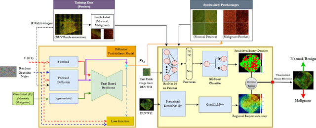
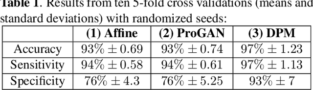

Data limitation is a significant challenge in applying deep learning to medical images. Recently, the diffusion probabilistic model (DPM) has shown the potential to generate high-quality images by converting Gaussian random noise into realistic images. In this paper, we apply the DPM to augment the deep ultraviolet fluorescence (DUV) image dataset with an aim to improve breast cancer classification for intraoperative margin assessment. For classification, we divide the whole surface DUV image into small patches and extract convolutional features for each patch by utilizing the pre-trained ResNet. Then, we feed them into an XGBoost classifier for patch-level decisions and then fuse them with a regional importance map computed by Grad-CAM++ for whole surface-level prediction. Our experimental results show that augmenting the training dataset with the DPM significantly improves breast cancer detection performance in DUV images, increasing accuracy from 93% to 97%, compared to using Affine transformations and ProGAN.