 Add to Chrome
 Add to Firefox
 Add to Edge
CRIL-U-Net: Compact Ratio-Interaction Learning for Focal Cortical Dysplasia Segmentation from T1w and FLAIR MRI
Aug 04, 2026
Focal cortical dysplasia (FCD) type II is an important structural cause of drug-resistant focal epilepsy, but its small size, heterogeneous appearance, and subtle MRI characteristics make automated segmentation challenging. Conventional multimodal networks commonly concatenate T1-weighted (T1w) and fluid-attenuated inversion recovery (FLAIR) images, requiring subsequent layers to learn useful cross-modal relationships implicitly. We propose CRIL-U-Net, a 3D U-Net incorporating a Compact Ratio-Interaction Learning module that combines local spatial features, voxel-wise cross-modal mixing, and bidirectional ratio-inspired interactions. CRIL-U-Net was compared with a conventional 3D U-Net and an input self-attention U-Net using five-fold cross-validation on 85 FCD subjects and 25 healthy controls. Each architecture was trained independently using Dice-binary cross-entropy (Dice-BCE) and Focal Tversky-Focal (FTF) losses. With FTF, CRIL-U-Net achieved the highest mean Dice score (0.196 +/- 0.262), compared with 0.136 +/- 0.224 for the U-Net and 0.135 +/- 0.214 for the attention comparator. It produced nonzero lesion overlap in 44 of 85 cases, compared with 36 for the U-Net. Under FTF, CRIL-U-Net significantly outperformed both comparison architectures after false-discovery-rate correction. These findings suggest that compact cross-modal representation learning can improve FCD segmentation within a controlled U-Net setting when combined with an imbalance-aware objective, although the remaining zero-overlap rate of 48.2% highlights the need for further validation and methodological development.

Benchmarking MRI Representations for Deep Learning-Based Focal Cortical Dysplasia Segmentation
Jul 17, 2026Focal cortical dysplasia (FCD) is one of the leading structural causes of drug-resistant focal epilepsy, yet its subtle and heterogeneous imaging characteristics make accurate identification and delineation challenging on conventional magnetic resonance imaging (MRI). Although T1-weighted (T1w) and fluid-attenuated inversion recovery (FLAIR) images are routinely acquired for presurgical evaluation, the contribution of different MRI representations to deep learning-based FCD segmentation remains poorly understood. In this study, we present a systematic benchmark of MRI representations for automated FCD segmentation using the nnU-Net framework. A publicly available presurgical MRI dataset comprising 85 FCD subjects and 25 healthy controls was used to evaluate eight input configurations, including conventional MRI contrasts (T1w and FLAIR), ratio-derived representations, and their multimodal combinations. To isolate the effect of MRI representation, all experiments employed identical preprocessing, network architecture, optimization strategy, and five-fold cross-validation. Among the evaluated single-modality representations, FLAIR achieved the strongest overall performance, whereas ratio-derived representations alone were insufficient for reliable identification of subtle FCD. Incorporating ratio-derived representations with conventional T1w and FLAIR images consistently improved lesion delineation, with the four-channel multimodal configuration achieving the highest overall Dice score (0.376), representing a 5.0% relative improvement over the conventional T1w+FLAIR representation. These findings demonstrate that MRI representation design is an important yet underexplored component of deep learning-based FCD segmentation and should be optimized alongside network architecture.
NeuroMorphix: A Novel Brain MRI Asymmetry-specific Feature Construction Approach For Seizure Recurrence Prediction
Apr 16, 2024
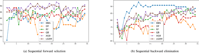
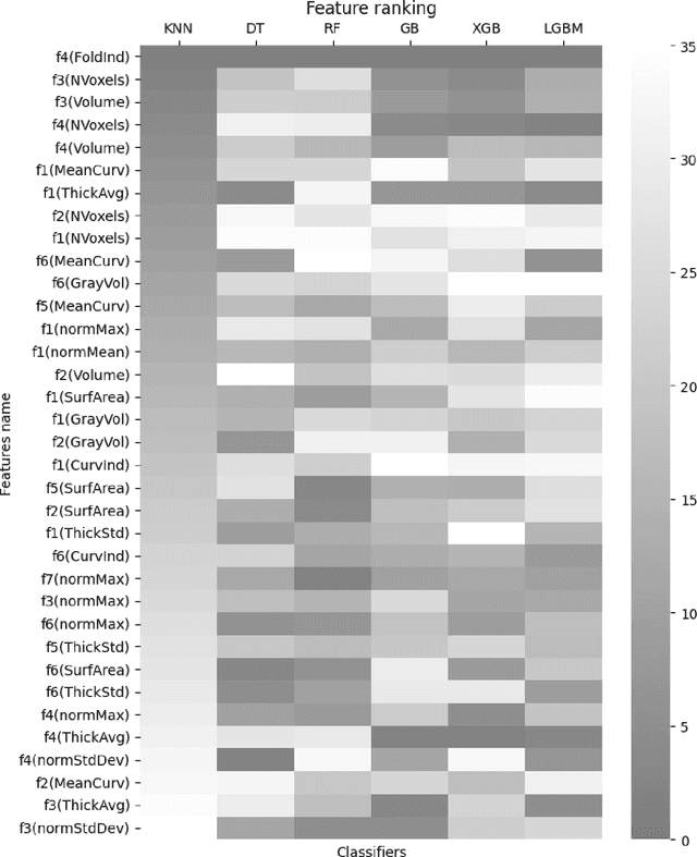

Seizure recurrence is an important concern after an initial unprovoked seizure; without drug treatment, it occurs within 2 years in 40-50% of cases. The decision to treat currently relies on predictors of seizure recurrence risk that are inaccurate, resulting in unnecessary, possibly harmful, treatment in some patients and potentially preventable seizures in others. Because of the link between brain lesions and seizure recurrence, we developed a recurrence prediction tool using machine learning and clinical 3T brain MRI. We developed NeuroMorphix, a feature construction approach based on MRI brain anatomy. Each of seven NeuroMorphix features measures the absolute or relative difference between corresponding regions in each cerebral hemisphere. FreeSurfer was used to segment brain regions and to generate values for morphometric parameters (8 for each cortical region and 5 for each subcortical region). The parameters were then mapped to whole brain NeuroMorphix features, yielding a total of 91 features per subject. Features were generated for a first seizure patient cohort (n = 169) categorised into seizure recurrence and non-recurrence subgroups. State-of-the-art classification algorithms were trained and tested using NeuroMorphix features to predict seizure recurrence. Classification models using the top 5 features, ranked by sequential forward selection, demonstrated excellent performance in predicting seizure recurrence, with area under the ROC curve of 88-93%, accuracy of 83-89%, and F1 score of 83-90%. Highly ranked features aligned with structural alterations known to be associated with epilepsy. This study highlights the potential for targeted, data-driven approaches to aid clinical decision-making in brain disorders.
RCCNet: An Efficient Convolutional Neural Network for Histological Routine Colon Cancer Nuclei Classification
Oct 20, 2018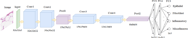
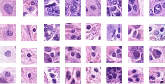
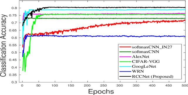

Efficient and precise classification of histological cell nuclei is of utmost importance due to its potential applications in the field of medical image analysis. It would facilitate the medical practitioners to better understand and explore various factors for cancer treatment. The classification of histological cell nuclei is a challenging task due to the cellular heterogeneity. This paper proposes an efficient Convolutional Neural Network (CNN) based architecture for classification of histological routine colon cancer nuclei named as RCCNet. The main objective of this network is to keep the CNN model as simple as possible. The proposed RCCNet model consists of only 1,512,868 learnable parameters which are significantly less compared to the popular CNN models such as AlexNet, CIFARVGG, GoogLeNet, and WRN. The experiments are conducted over publicly available routine colon cancer histological dataset "CRCHistoPhenotypes". The results of the proposed RCCNet model are compared with five state-of-the-art CNN models in terms of the accuracy, weighted average F1 score and training time. The proposed method has achieved a classification accuracy of 80.61% and 0.7887 weighted average F1 score. The proposed RCCNet is more efficient and generalized terms of the training time and data over-fitting, respectively.