 Add to Chrome
 Add to Firefox
 Add to Edge
CRIL-U-Net: Compact Ratio-Interaction Learning for Focal Cortical Dysplasia Segmentation from T1w and FLAIR MRI
Aug 04, 2026
Focal cortical dysplasia (FCD) type II is an important structural cause of drug-resistant focal epilepsy, but its small size, heterogeneous appearance, and subtle MRI characteristics make automated segmentation challenging. Conventional multimodal networks commonly concatenate T1-weighted (T1w) and fluid-attenuated inversion recovery (FLAIR) images, requiring subsequent layers to learn useful cross-modal relationships implicitly. We propose CRIL-U-Net, a 3D U-Net incorporating a Compact Ratio-Interaction Learning module that combines local spatial features, voxel-wise cross-modal mixing, and bidirectional ratio-inspired interactions. CRIL-U-Net was compared with a conventional 3D U-Net and an input self-attention U-Net using five-fold cross-validation on 85 FCD subjects and 25 healthy controls. Each architecture was trained independently using Dice-binary cross-entropy (Dice-BCE) and Focal Tversky-Focal (FTF) losses. With FTF, CRIL-U-Net achieved the highest mean Dice score (0.196 +/- 0.262), compared with 0.136 +/- 0.224 for the U-Net and 0.135 +/- 0.214 for the attention comparator. It produced nonzero lesion overlap in 44 of 85 cases, compared with 36 for the U-Net. Under FTF, CRIL-U-Net significantly outperformed both comparison architectures after false-discovery-rate correction. These findings suggest that compact cross-modal representation learning can improve FCD segmentation within a controlled U-Net setting when combined with an imbalance-aware objective, although the remaining zero-overlap rate of 48.2% highlights the need for further validation and methodological development.

Benchmarking MRI Representations for Deep Learning-Based Focal Cortical Dysplasia Segmentation
Jul 17, 2026Focal cortical dysplasia (FCD) is one of the leading structural causes of drug-resistant focal epilepsy, yet its subtle and heterogeneous imaging characteristics make accurate identification and delineation challenging on conventional magnetic resonance imaging (MRI). Although T1-weighted (T1w) and fluid-attenuated inversion recovery (FLAIR) images are routinely acquired for presurgical evaluation, the contribution of different MRI representations to deep learning-based FCD segmentation remains poorly understood. In this study, we present a systematic benchmark of MRI representations for automated FCD segmentation using the nnU-Net framework. A publicly available presurgical MRI dataset comprising 85 FCD subjects and 25 healthy controls was used to evaluate eight input configurations, including conventional MRI contrasts (T1w and FLAIR), ratio-derived representations, and their multimodal combinations. To isolate the effect of MRI representation, all experiments employed identical preprocessing, network architecture, optimization strategy, and five-fold cross-validation. Among the evaluated single-modality representations, FLAIR achieved the strongest overall performance, whereas ratio-derived representations alone were insufficient for reliable identification of subtle FCD. Incorporating ratio-derived representations with conventional T1w and FLAIR images consistently improved lesion delineation, with the four-channel multimodal configuration achieving the highest overall Dice score (0.376), representing a 5.0% relative improvement over the conventional T1w+FLAIR representation. These findings demonstrate that MRI representation design is an important yet underexplored component of deep learning-based FCD segmentation and should be optimized alongside network architecture.
Uncertainty-Guided Cross Attention Ensemble Mean Teacher for Semi-supervised Medical Image Segmentation
Dec 19, 2024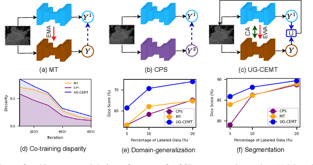
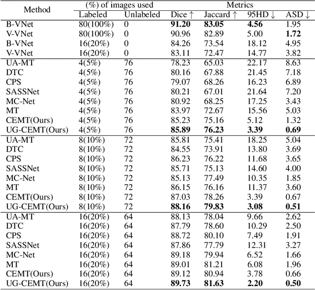
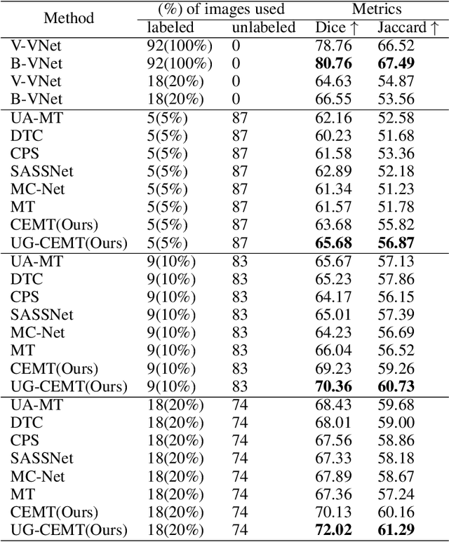
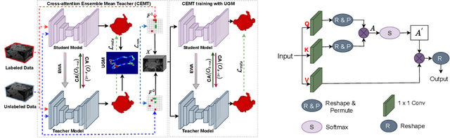
This work proposes a novel framework, Uncertainty-Guided Cross Attention Ensemble Mean Teacher (UG-CEMT), for achieving state-of-the-art performance in semi-supervised medical image segmentation. UG-CEMT leverages the strengths of co-training and knowledge distillation by combining a Cross-attention Ensemble Mean Teacher framework (CEMT) inspired by Vision Transformers (ViT) with uncertainty-guided consistency regularization and Sharpness-Aware Minimization emphasizing uncertainty. UG-CEMT improves semi-supervised performance while maintaining a consistent network architecture and task setting by fostering high disparity between sub-networks. Experiments demonstrate significant advantages over existing methods like Mean Teacher and Cross-pseudo Supervision in terms of disparity, domain generalization, and medical image segmentation performance. UG-CEMT achieves state-of-the-art results on multi-center prostate MRI and cardiac MRI datasets, where object segmentation is particularly challenging. Our results show that using only 10\% labeled data, UG-CEMT approaches the performance of fully supervised methods, demonstrating its effectiveness in exploiting unlabeled data for robust medical image segmentation. The code is publicly available at \url{https://github.com/Meghnak13/UG-CEMT}