 Add to Chrome
 Add to Firefox
 Add to Edge
Multimodal Ordinal Modeling of Alzheimer's Disease Severity Using Structural MRI and Clinical Data
Jun 10, 2026Neurodegenerative diseases such as Alzheimer's disease (AD) require accurate and scalable tools for assessing disease severity, yet current clinical staging remains time-intensive and prone to variability. We propose an attention-enhanced multimodal machine learning framework with ordinal regression for automated and interpretable AD severity staging. The framework integrates T1-weighted MRI with demographic and genetic variables and compares unimodal and multimodal architectures using ordinal and non-ordinal prediction heads. Models were trained and validated using cohort-stratified splits derived from the ADNI, AIBL, and NIFD datasets. A strictly held-out test set was constructed using subjects excluded from all training, validation, preprocessing, and hyperparameter tuning procedures, with subject-level splitting employed throughout to prevent data leakage. Among unimodal approaches, the T1-weighted MRI model achieved slightly higher adjacent-stage accuracy (0.963) and agreement with clinical staging (QWK 0.444) than the tabular model (QWK 0.433). Integrating imaging, demographic, and genetic information improved overall performance. The multimodal non-ordinal baseline achieved the lowest prediction error (MAE 0.340), whereas the ordinal multimodal model achieved the highest adjacent-stage accuracy (0.970) and strongest agreement with clinical staging (QWK 0.549). These findings indicate that ordinal formulations better capture the ordered structure of the CDR scale and yield predictions more consistent with clinical staging. Explainability analyses using Grad CAM++ and SHAP demonstrated anatomically and clinically plausible model behavior, supporting transparent decision-making. Overall, attention-based multimodal learning with ordinal regression represents a robust, interpretable, and scalable approach for automated AD severity staging and AI-assisted clinical decision support.
Improving 3D convolutional neural network comprehensibility via interactive visualization of relevance maps: Evaluation in Alzheimer's disease
Dec 18, 2020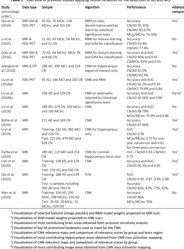
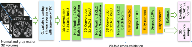
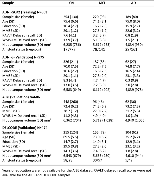
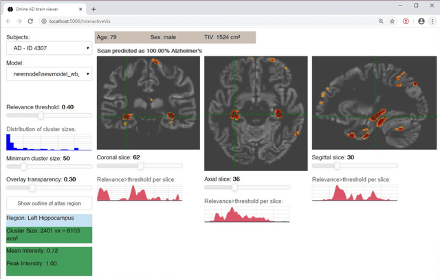
Although convolutional neural networks (CNN) achieve high diagnostic accuracy for detecting Alzheimer's disease (AD) dementia based on magnetic resonance imaging (MRI) scans, they are not yet applied in clinical routine. One important reason for this is a lack of model comprehensibility. Recently developed visualization methods for deriving CNN relevance maps may help to fill this gap. We investigated whether models with higher accuracy also rely more on discriminative brain regions predefined by prior knowledge. We trained a CNN for the detection of AD in N=663 T1-weighted MRI scans of patients with dementia and amnestic mild cognitive impairment (MCI) and verified the accuracy of the models via cross-validation and in three independent samples including N=1655 cases. We evaluated the association of relevance scores and hippocampus volume to validate the clinical utility of this approach. To improve model comprehensibility, we implemented an interactive visualization of 3D CNN relevance maps. Across three independent datasets, group separation showed high accuracy for AD dementia vs. controls (AUC$\geq$0.92) and moderate accuracy for MCI vs. controls (AUC$\approx$0.75). Relevance maps indicated that hippocampal atrophy was considered as the most informative factor for AD detection, with additional contributions from atrophy in other cortical and subcortical regions. Relevance scores within the hippocampus were highly correlated with hippocampal volumes (Pearson's r$\approx$-0.81). The relevance maps highlighted atrophy in regions that we had hypothesized a priori. This strengthens the comprehensibility of the CNN models, which were trained in a purely data-driven manner based on the scans and diagnosis labels. The high hippocampus relevance scores and high performance achieved in independent samples support the validity of the CNN models in the detection of AD-related MRI abnormalities.