 Add to Chrome
 Add to Firefox
 Add to Edge
BreastMammo and DenseMammo: Benchmarks for Mammography Domain Generalization
Aug 10, 2026
Breast density classification is a critical component of breast cancer risk assessment, yet AI models often struggle to generalize across clinical sites due to vendor-specific acquisition styles. In this work, we introduce two new datasets, BreastMammo and DenseMammo, to facilitate robust multi-view mammography research. We propose a domain generalization framework that utilizes a foreground-only histogram matching protocol to resolve the domain shift issue arising from disparate clinical sources. Internal evaluation using a 5-fold cross-validation protocol demonstrates the efficacy of our approach, with the Swin Transformer backbone achieving a peak AUC of 98.32% for density classification. External evaluation on the TNMammo and LUMINA datasets demonstrates that the proposed approach consistently reduces domain shift, significantly outperforming prominent domain generalization paradigms, including MixStyle and Discrete-Fourier-Transform-based frameworks.

SPARC-Rad: A Multimodal Benchmark Dataset and Evaluation Pipeline for Spatial and Anatomical Reasoning in Radiology Vision-Language Models
Jul 30, 2026Vision-language models (VLMs) are increasingly being evaluated for medical imaging, but many available benchmarks emphasize disease classification, report generation, or broad visual question answering rather than the spatial and anatomical reasoning required for radiology. We developed the Spatial Perception and Anatomical Reasoning in Clinical Radiology (SPARC-Rad) Benchmark, a manually curated multimodal benchmark dataset and evaluation pipeline for assessing these capabilities in radiology VLMs. SPARC-Rad includes 300 image-question pairs derived from healthy control imaging studies in The Cancer Imaging Archive (TCIA), spanning CT, MRI, and radiography across the abdomen, chest, breast, neuro, and musculoskeletal categories. Radiology trainees manually designed and annotated questions to evaluate anatomical identification, localization, laterality, regional recognition, device identification, and inter-structure spatial relationships. The evaluation pipeline supports standardized prompting, structured output collection, response normalization, LLM-as-judge grading, human quality review, binary correctness scoring, and subgroup analysis by modality, anatomy, and reasoning type. SPARC-Rad provides a reusable framework for evaluating whether VLMs can provide reasoning for radiologic anatomy as a spatial system, supporting future model development, failure-mode analysis, and pre-deployment assessment.
Mammographic Breast Positioning Assessment via Deep Learning
Jul 15, 2024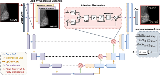
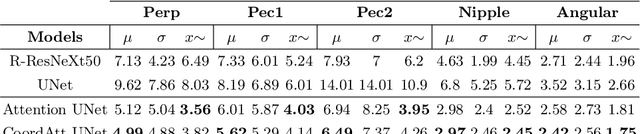
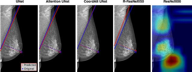
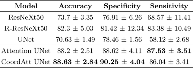
Breast cancer remains a leading cause of cancer-related deaths among women worldwide, with mammography screening as the most effective method for the early detection. Ensuring proper positioning in mammography is critical, as poor positioning can lead to diagnostic errors, increased patient stress, and higher costs due to recalls. Despite advancements in deep learning (DL) for breast cancer diagnostics, limited focus has been given to evaluating mammography positioning. This paper introduces a novel DL methodology to quantitatively assess mammogram positioning quality, specifically in mediolateral oblique (MLO) views using attention and coordinate convolution modules. Our method identifies key anatomical landmarks, such as the nipple and pectoralis muscle, and automatically draws a posterior nipple line (PNL), offering robust and inherently explainable alternative to well-known classification and regression-based approaches. We compare the performance of proposed methodology with various regression and classification-based models. The CoordAtt UNet model achieved the highest accuracy of 88.63% $\pm$ 2.84 and specificity of 90.25% $\pm$ 4.04, along with a noteworthy sensitivity of 86.04% $\pm$ 3.41. In landmark detection, the same model also recorded the lowest mean errors in key anatomical points and the smallest angular error of 2.42 degrees. Our results indicate that models incorporating attention mechanisms and CoordConv module increase the accuracy in classifying breast positioning quality and detecting anatomical landmarks. Furthermore, we make the labels and source codes available to the community to initiate an open research area for mammography, accessible at https://github.com/tanyelai/deep-breast-positioning.
Prostate Lesion Estimation using Prostate Masks from Biparametric MRI
Jan 11, 2023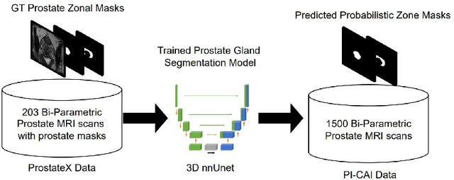
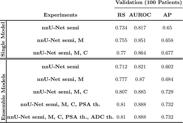
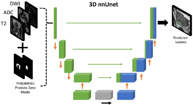
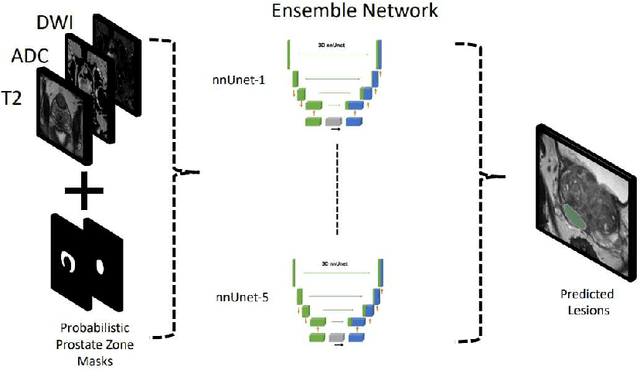
Biparametric MRI has emerged as an alternative to multiparametric prostate MRI, which eliminates the need for the potential harms to the patient due to the contrast medium. One major issue with biparametric MRI is difficulty to detect clinically significant prostate cancer (csPCA). Deep learning algorithms have emerged as an alternative solution to detect csPCA in cohort studies. We present a workflow which predicts csPCA on biparametric prostate MRI PI-CAI 2022 Challenge with over 10,000 carefully-curated prostate MRI exams. We propose to to segment the prostate gland first to the central gland (transition + central zone) and the peripheral gland. Then we utilize these predcitions in combination with T2, ADC and DWI images to train an ensemble nnU-Net model. Finally, we utilize clinical indices PSA and ADC intensity distributions of lesion regions to reduce the false positives. Our method achieves top results on open-validation stage with a AUROC of 0.888 and AP of 0.732.