 Add to Chrome
 Add to Firefox
 Add to Edge
Comparing Human Gaze and Vision-Language Model Attention in Safety-Relevant Environments
Jun 13, 2026Human visual attention plays an important role in how people perceive and respond to environments containing potential risks. This study investigates whether large vision-language models can identify the same regions of a scene that attract human attention in safety-relevant environments. Eye-tracking data were collected from ten participants viewing 33 scene images representing environments with varying levels of potential risk using Pupil Invisible wearable glasses. Gaze coordinates were mapped onto stimulus images to generate population-averaged human gaze heatmaps. In parallel, GPT-4o was prompted through the OpenAI Vision Application Programming Interface (API) to generate spatial predictions of visual attention, which were converted into saliency maps for comparison with human gaze patterns. Spatial alignment between human gaze heatmaps and model-generated saliency maps was evaluated using four complementary metrics: Pearson correlation (r = 0.515 +- 0.117), Normalised Scanpath Saliency (NSS = 0.988 +- 0.323), Kullback-Leibler divergence (KL = 1.766 +- 0.844), and Area Under the Receiver Operating Characteristic Curve using the Judd formulation (AUC-Judd = 0.806 +- 0.076). A cross-model comparison with Gemini Pro, Gemini Flash, and Claude showed that all models exceeded the AUC-Judd chance baseline of 0.5 and achieved positive NSS scores. Gemini Pro demonstrated the strongest spatial localisation according to three of the four metrics, whereas GPT-4o produced the closest distributional match to human attention as measured by KL divergence. These findings suggest that large vision-language models can identify regions that broadly correspond to where humans direct visual attention in safety-relevant scenes without requiring eye-tracking training data. The results highlight the potential of vision-language models as a scalable tool for approximating human attentional patterns.
Validating Vision Transformers for Otoscopy: Performance and Data-Leakage Effects
Nov 06, 2025


This study evaluates the efficacy of vision transformer models, specifically Swin transformers, in enhancing the diagnostic accuracy of ear diseases compared to traditional convolutional neural networks. With a reported 27% misdiagnosis rate among specialist otolaryngologists, improving diagnostic accuracy is crucial. The research utilised a real-world dataset from the Department of Otolaryngology at the Clinical Hospital of the Universidad de Chile, comprising otoscopic videos of ear examinations depicting various middle and external ear conditions. Frames were selected based on the Laplacian and Shannon entropy thresholds, with blank frames removed. Initially, Swin v1 and Swin v2 transformer models achieved accuracies of 100% and 99.1%, respectively, marginally outperforming the ResNet model (99.5%). These results surpassed metrics reported in related studies. However, the evaluation uncovered a critical data leakage issue in the preprocessing step, affecting both this study and related research using the same raw dataset. After mitigating the data leakage, model performance decreased significantly. Corrected accuracies were 83% for both Swin v1 and Swin v2, and 82% for the ResNet model. This finding highlights the importance of rigorous data handling in machine learning studies, especially in medical applications. The findings indicate that while vision transformers show promise, it is essential to find an optimal balance between the benefits of advanced model architectures and those derived from effective data preprocessing. This balance is key to developing a reliable machine learning model for diagnosing ear diseases.
Deep Learning-Assisted Co-registration of Full-Spectral Autofluorescence Lifetime Microscopic Images with H&E-Stained Histology Images
Feb 15, 2022
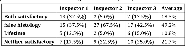
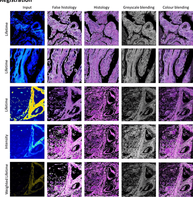
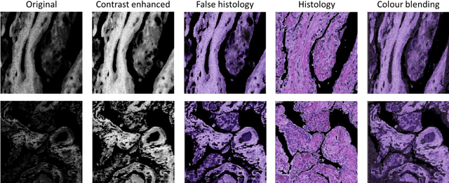
Autofluorescence lifetime images reveal unique characteristics of endogenous fluorescence in biological samples. Comprehensive understanding and clinical diagnosis rely on co-registration with the gold standard, histology images, which is extremely challenging due to the difference of both images. Here, we show an unsupervised image-to-image translation network that significantly improves the success of the co-registration using a conventional optimisation-based regression network, applicable to autofluorescence lifetime images at different emission wavelengths. A preliminary blind comparison by experienced researchers shows the superiority of our method on co-registration. The results also indicate that the approach is applicable to various image formats, like fluorescence intensity images. With the registration, stitching outcomes illustrate the distinct differences of the spectral lifetime across an unstained tissue, enabling macro-level rapid visual identification of lung cancer and cellular-level characterisation of cell variants and common types. The approach could be effortlessly extended to lifetime images beyond this range and other staining technologies.