 Add to Chrome
 Add to Firefox
 Add to Edge
Subject-independent Classification of Meditative State from the Resting State using EEG
Apr 25, 2025


While it is beneficial to objectively determine whether a subject is meditating, most research in the literature reports good results only in a subject-dependent manner. This study aims to distinguish the modified state of consciousness experienced during Rajyoga meditation from the resting state of the brain in a subject-independent manner using EEG data. Three architectures have been proposed and evaluated: The CSP-LDA Architecture utilizes common spatial pattern (CSP) for feature extraction and linear discriminant analysis (LDA) for classification. The CSP-LDA-LSTM Architecture employs CSP for feature extraction, LDA for dimensionality reduction, and long short-term memory (LSTM) networks for classification, modeling the binary classification problem as a sequence learning problem. The SVD-NN Architecture uses singular value decomposition (SVD) to select the most relevant components of the EEG signals and a shallow neural network (NN) for classification. The CSP-LDA-LSTM architecture gives the best performance with 98.2% accuracy for intra-subject classification. The SVD-NN architecture provides significant performance with 96.4\% accuracy for inter-subject classification. This is comparable to the best-reported accuracies in the literature for intra-subject classification. Both architectures are capable of capturing subject-invariant EEG features for effectively classifying the meditative state from the resting state. The high intra-subject and inter-subject classification accuracies indicate these systems' robustness and their ability to generalize across different subjects.
A Pipelined Memristive Neural Network Analog-to-Digital Converter
Jun 04, 2024


With the advent of high-speed, high-precision, and low-power mixed-signal systems, there is an ever-growing demand for accurate, fast, and energy-efficient analog-to-digital (ADCs) and digital-to-analog converters (DACs). Unfortunately, with the downscaling of CMOS technology, modern ADCs trade off speed, power and accuracy. Recently, memristive neuromorphic architectures of four-bit ADC/DAC have been proposed. Such converters can be trained in real-time using machine learning algorithms, to break through the speedpower-accuracy trade-off while optimizing the conversion performance for different applications. However, scaling such architectures above four bits is challenging. This paper proposes a scalable and modular neural network ADC architecture based on a pipeline of four-bit converters, preserving their inherent advantages in application reconfiguration, mismatch selfcalibration, noise tolerance, and power optimization, while approaching higher resolution and throughput in penalty of latency. SPICE evaluation shows that an 8-bit pipelined ADC achieves 0.18 LSB INL, 0.20 LSB DNL, 7.6 ENOB, and 0.97 fJ/conv FOM. This work presents a significant step towards the realization of large-scale neuromorphic data converters.
Functional Connectivity Methods for EEG-based Biometrics on a Large, Heterogeneous Dataset
Jun 03, 2022
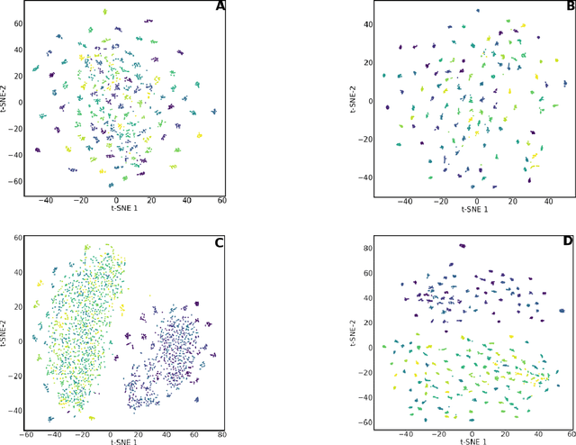
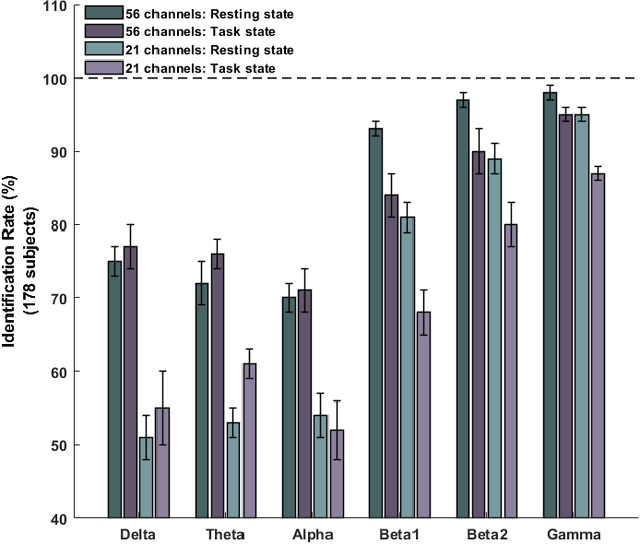
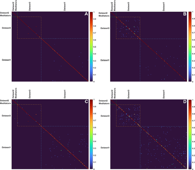
This study examines the utility of functional connectivity (FC) and graph-based (GB) measures with a support vector machine classifier for use in electroencephalogram (EEG) based biometrics. Although FC-based features have been used in biometric applications, studies assessing the identification algorithms on heterogeneous and large datasets are scarce. This work investigates the performance of FC and GB metrics on a dataset of 184 subjects formed by pooling three datasets recorded under different protocols and acquisition systems. The results demonstrate the higher discriminatory power of FC than GB metrics. The identification accuracy increases with higher frequency EEG bands, indicating the enhanced uniqueness of the neural signatures in beta and gamma bands. Using all the 56 EEG channels common to the three databases, the best identification accuracy of 97.4% is obtained using phase-locking value (PLV) based measures extracted from the gamma frequency band. Further, we investigate the effect of the length of the analysis epoch to determine the data acquisition time required to obtain satisfactory identification accuracy. When the number of channels is reduced to 21 from 56, there is a marginal reduction of 2.4% only in the identification accuracy using PLV features in the gamma band. Additional experiments have been conducted to study the effect of the cognitive state of the subject and mismatched train/test conditions on the performance of the system.
An Improved EEG Acquisition Protocol Facilitates Localized Neural Activation
Mar 13, 2020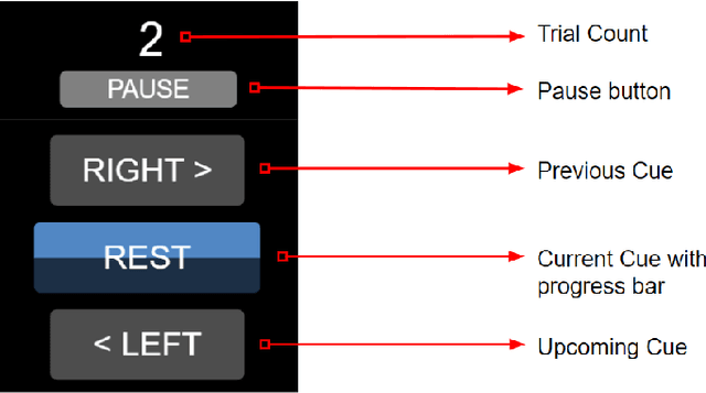
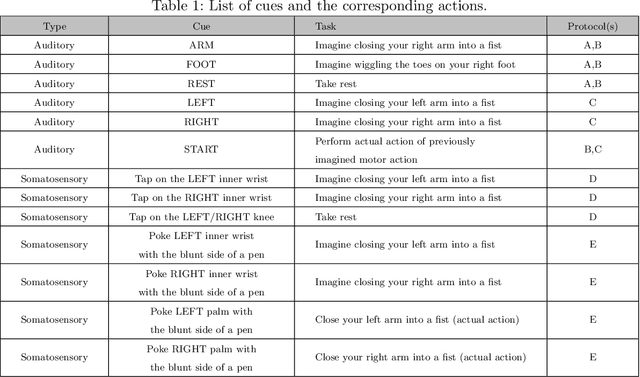
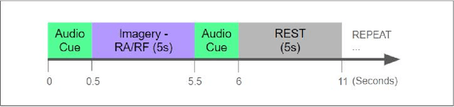
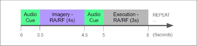
This work proposes improvements in the electroencephalogram (EEG) recording protocols for motor imagery through the introduction of actual motor movement and/or somatosensory cues. The results obtained demonstrate the advantage of requiring the subjects to perform motor actions following the trials of imagery. By introducing motor actions in the protocol, the subjects are able to perform actual motor planning, rather than just visualizing the motor movement, thus greatly improving the ease with which the motor movements can be imagined. This study also probes the added advantage of administering somatosensory cues in the subject, as opposed to the conventional auditory/visual cues. These changes in the protocol show promise in terms of the aptness of the spatial filters obtained on the data, on application of the well-known common spatial pattern (CSP) algorithms. The regions highlighted by the spatial filters are more localized and consistent across the subjects when the protocol is augmented with somatosensory stimuli. Hence, we suggest that this may prove to be a better EEG acquisition protocol for detecting brain activation in response to intended motor commands in (clinically) paralyzed/locked-in patients.
Semi-Automatic Segmentation of Autosomal Dominant Polycystic Kidneys using Random Forests
Oct 23, 2015
This paper presents a method for 3D segmentation of kidneys from patients with autosomal dominant polycystic kidney disease (ADPKD) and severe renal insufficiency, using computed tomography (CT) data. ADPKD severely alters the shape of the kidneys due to non-uniform formation of cysts. As a consequence, fully automatic segmentation of such kidneys is very challenging. We present a segmentation method with minimal user interaction based on a random forest classifier. One of the major novelties of the proposed approach is the usage of geodesic distance volumes as additional source of information. These volumes contain the intensity weighted distance to a manual outline of the respective kidney in only one slice (for each kidney) of the CT volume. We evaluate our method qualitatively and quantitatively on 55 CT acquisitions using ground truth annotations from clinical experts.