 Add to Chrome
 Add to Firefox
 Add to Edge
Asymmetry Disentanglement Network for Interpretable Acute Ischemic Stroke Infarct Segmentation in Non-Contrast CT Scans
Jun 30, 2022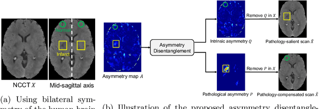
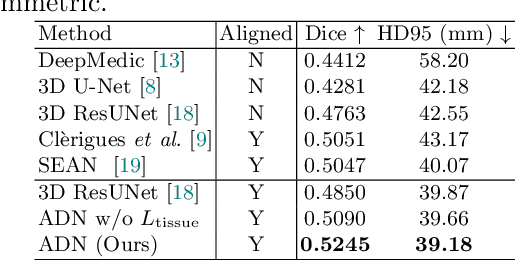
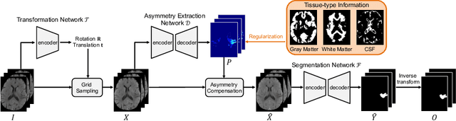
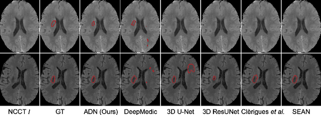
Accurate infarct segmentation in non-contrast CT (NCCT) images is a crucial step toward computer-aided acute ischemic stroke (AIS) assessment. In clinical practice, bilateral symmetric comparison of brain hemispheres is usually used to locate pathological abnormalities. Recent research has explored asymmetries to assist with AIS segmentation. However, most previous symmetry-based work mixed different types of asymmetries when evaluating their contribution to AIS. In this paper, we propose a novel Asymmetry Disentanglement Network (ADN) to automatically separate pathological asymmetries and intrinsic anatomical asymmetries in NCCTs for more effective and interpretable AIS segmentation. ADN first performs asymmetry disentanglement based on input NCCTs, which produces different types of 3D asymmetry maps. Then a synthetic, intrinsic-asymmetry-compensated and pathology-asymmetry-salient NCCT volume is generated and later used as input to a segmentation network. The training of ADN incorporates domain knowledge and adopts a tissue-type aware regularization loss function to encourage clinically-meaningful pathological asymmetry extraction. Coupled with an unsupervised 3D transformation network, ADN achieves state-of-the-art AIS segmentation performance on a public NCCT dataset. In addition to the superior performance, we believe the learned clinically-interpretable asymmetry maps can also provide insights towards a better understanding of AIS assessment. Our code is available at https://github.com/nihaomiao/MICCAI22_ADN.
Patcher: Patch Transformers with Mixture of Experts for Precise Medical Image Segmentation
Jun 03, 2022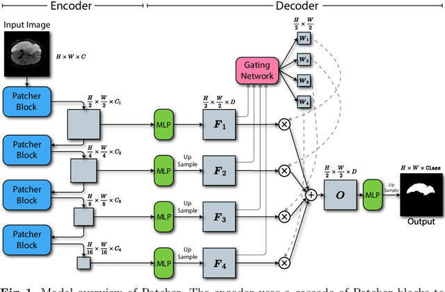
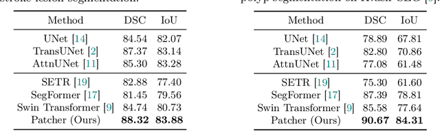
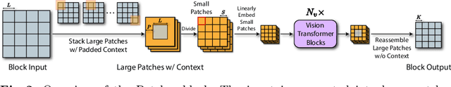
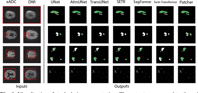
We present a new encoder-decoder Vision Transformer architecture, Patcher, for medical image segmentation. Unlike standard Vision Transformers, it employs Patcher blocks that segment an image into large patches, each of which is further divided into small patches. Transformers are applied to the small patches within a large patch, which constrains the receptive field of each pixel. We intentionally make the large patches overlap to enhance intra-patch communication. The encoder employs a cascade of Patcher blocks with increasing receptive fields to extract features from local to global levels. This design allows Patcher to benefit from both the coarse-to-fine feature extraction common in CNNs and the superior spatial relationship modeling of Transformers. We also propose a new mixture-of-experts (MoE) based decoder, which treats the feature maps from the encoder as experts and selects a suitable set of expert features to predict the label for each pixel. The use of MoE enables better specializations of the expert features and reduces interference between them during inference. Extensive experiments demonstrate that Patcher outperforms state-of-the-art Transformer- and CNN-based approaches significantly on stroke lesion segmentation and polyp segmentation. Code for Patcher will be released with publication to facilitate future research.
DeepStroke: An Efficient Stroke Screening Framework for Emergency Rooms with Multimodal Adversarial Deep Learning
Sep 24, 2021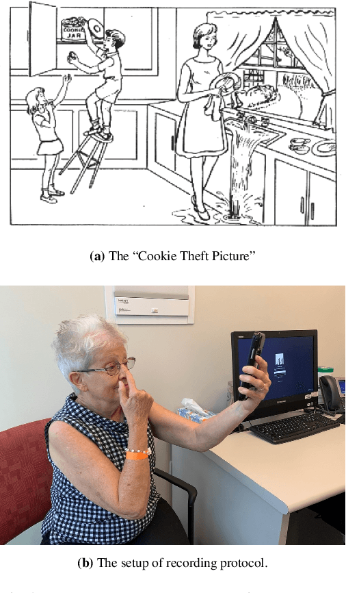
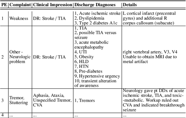
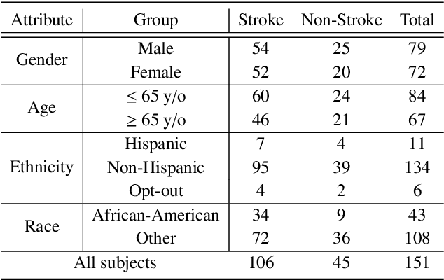
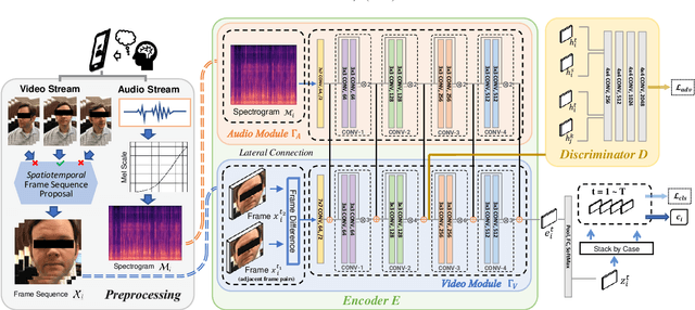
In an emergency room (ER) setting, the diagnosis of stroke is a common challenge. Due to excessive execution time and cost, an MRI scan is usually not available in the ER. Clinical tests are commonly referred to in stroke screening, but neurologists may not be immediately available. We propose a novel multimodal deep learning framework, DeepStroke, to achieve computer-aided stroke presence assessment by recognizing the patterns of facial motion incoordination and speech inability for patients with suspicion of stroke in an acute setting. Our proposed DeepStroke takes video data for local facial paralysis detection and audio data for global speech disorder analysis. It further leverages a multi-modal lateral fusion to combine the low- and high-level features and provides mutual regularization for joint training. A novel adversarial training loss is also introduced to obtain identity-independent and stroke-discriminative features. Experiments on our video-audio dataset with actual ER patients show that the proposed approach outperforms state-of-the-art models and achieves better performance than ER doctors, attaining a 6.60% higher sensitivity and maintaining 4.62% higher accuracy when specificity is aligned. Meanwhile, each assessment can be completed in less than 6 minutes, demonstrating the framework's great potential for clinical implementation.
LambdaUNet: 2.5D Stroke Lesion Segmentation of Diffusion-weighted MR Images
Apr 28, 2021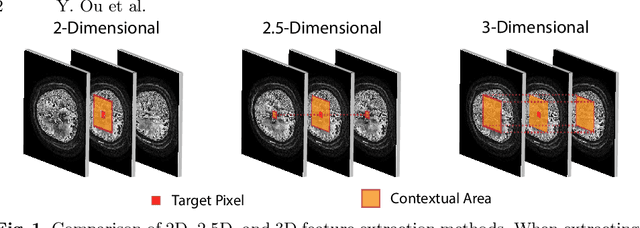
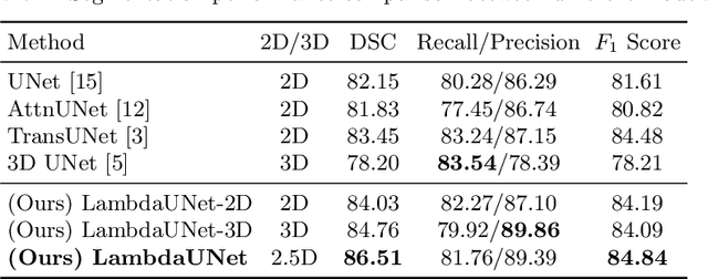
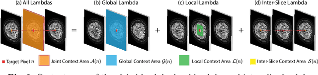
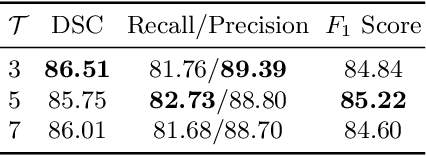
Diffusion-weighted (DW) magnetic resonance imaging is essential for the diagnosis and treatment of ischemic stroke. DW images (DWIs) are usually acquired in multi-slice settings where lesion areas in two consecutive 2D slices are highly discontinuous due to large slice thickness and sometimes even slice gaps. Therefore, although DWIs contain rich 3D information, they cannot be treated as regular 3D or 2D images. Instead, DWIs are somewhere in-between (or 2.5D) due to the volumetric nature but inter-slice discontinuities. Thus, it is not ideal to apply most existing segmentation methods as they are designed for either 2D or 3D images. To tackle this problem, we propose a new neural network architecture tailored for segmenting highly-discontinuous 2.5D data such as DWIs. Our network, termed LambdaUNet, extends UNet by replacing convolutional layers with our proposed Lambda+ layers. In particular, Lambda+ layers transform both intra-slice and inter-slice context around a pixel into linear functions, called lambdas, which are then applied to the pixel to produce informative 2.5D features. LambdaUNet is simple yet effective in combining sparse inter-slice information from adjacent slices while also capturing dense contextual features within a single slice. Experiments on a unique clinical dataset demonstrate that LambdaUNet outperforms existing 3D/2D image segmentation methods including recent variants of UNet. Code for LambdaUNet will be released with the publication to facilitate future research.