 Add to Chrome
 Add to Firefox
 Add to Edge
PolypVision: A Three-Stage Hierarchical Deep Learning Framework for Classification and Segmentation of Colorectal Polyps
Aug 11, 2026
Colorectal cancer (CRC) remains one of the leading causes of cancer-related mortality worldwide, predominantly arising from precancerous polyps. Accurate detection, segmentation, and endoscopic and histological classification of colorectal polyps are crucial for timely clinical intervention. In this study, we present PolypVision, a three-stage hierarchical deep learning framework that sequentially performs: (Stage 1) binary classification of polyps as adenomatous or hyperplastic, with simultaneous Paris and JNet classification, using EfficientNetV2-M with Focal Loss; (Stage 2) polyp segmentation with recommended resection method using a UNet++ decoder with the Stage 1 backbone as encoder, optimized with Dice and BCE losses; and (Stage 3) adenoma subtype classification (tubular, tubulovillous, villous) using EfficientNetV2-M with transfer learning from Stage 2. Evaluated on three public datasets -- PolypGen, Kvasir-SEG, and CVC-ClinicDB -- PolypVision achieves an AUC of approximately 0.99 for frame classification and a detection mAP@50 of 94.4% on Kvasir-SEG, outperforming or matching state-of-the-art methods. Gradient-weighted Class Activation Maps (Grad-CAM) confirm that the model attends to clinically relevant lesion features. The framework is device-independent, operating across diverse endoscopic imaging systems without hardware-specific adaptation. These results demonstrate that a hierarchical, transfer-learning-driven pipeline with task-specific loss functions offers a robust, device-independent, and clinically meaningful approach to automated colorectal polyp analysis. PolypVision is freely available as a web application at https://polypvision.com, a DataBioX initiative, with a free usage tier open to all users.

CRC-HGD: A Histopathological Image Dataset for Grading Colorectal Cancer
Jul 14, 2026Colorectal cancer (CRC) is the third most common cancer worldwide and the second leading cause of cancer-related deaths globally, with approximately 1,926,425 new cases and 904,019 deaths reported in 2022. Accurate histologic grading plays a critical role in prognosis and treatment planning for colorectal adenocarcinoma. In recent years, artificial intelligence and its subcategories, including machine learning and deep learning, have been increasingly employed for automated cancer detection and classification. An appropriate and well-organized dataset is the essential first step to achieve this goal. This paper introduces CRC-HGD, a histopathological microscopy image dataset of 1,914 images obtained from 214 colorectal adenocarcinoma patients (Grade I: 106, Grade II: 75, Grade III: 33). The specimens are H&E-stained colorectal tissue sections acquired at the Poursina Hakim Research Center of Isfahan University of Medical Sciences, Iran, diagnosed between 2014 and 2019, and graded according to the World Health Organization (WHO) criteria into three grades: well-differentiated (Grade I), moderately differentiated (Grade II), and poorly differentiated (Grade III). For each specimen, four magnification levels are provided: 4x, 10x, 20x, and 40x. The dataset is accessible via Mendeley Data (https://doi.org/10.17632/yfp5sfj47m.4) and at http://databiox.com, where the latest version is also available. The distinctive feature of this dataset is the provision of labeled specimens across all three differentiation grades at multiple magnification levels, enabling comprehensive computational analysis of colorectal cancer grading.
GIM-ENDO: A Multimodal Endoscopic Image and Video Dataset for Gastric Intestinal Metaplasia Morphology and Pathology
Jun 18, 2026Gastric intestinal metaplasia (GIM) is a precursor lesion to gastric dysplasia and adenocarcinoma whose early detection is crucial for intervening in the carcinogenesis cascade. Artificial intelligence (AI) holds considerable promise for real-time endoscopic detection and characterization of GIM. However, development of reliable AI models has been constrained by the absence of publicly available, histopathologically validated datasets that combine detailed endoscopic annotations, histological subtype (complete and incomplete), standardized grading systems, and normal mucosal patterns. GIM-ENDO was designed to fill this gap. The dataset comprises demographic data, endoscopic findings, histopathological results, and H. pylori status acquired using the Olympus EVIS X1 system with white-light endoscopy (WLE) and image-enhanced endoscopy (IEE), including narrow-band imaging (NBI) and magnifying NBI (M-NBI), along with images and video clips from 24 patients (22 GIM-positive, 2 normal controls). Annotations cover six primary IEE endoscopic signs -- light blue crest (LBC), marginal turbid band (MTB), white opaque substance (WOS), TV pattern (Fusion), atrophy, and map-like erythema (MLE) -- plus two additional endoscopic findings (AHP and GA) recorded where present. GIM subtypes (complete and incomplete) are annotated for all GIM-positive cases; OLGA and OLGIM staging are provided where complete histological sampling was available. The dataset is publicly accessible at https://doi.org/10.5281/zenodo.20707267. For the latest updates and further information regarding this dataset, readers are referred to the DataBioX website: https://databiox.com A short version of this work has been submitted to MICCAI 2026 Open Data Track.
A Review on the Applications of Machine Learning for Tinnitus Diagnosis Using EEG Signals
Oct 28, 2023
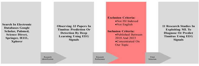
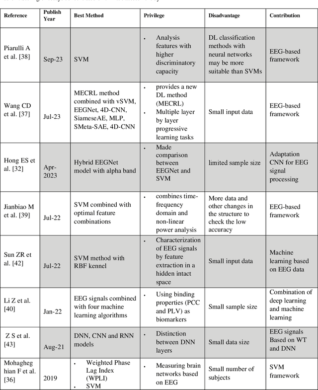
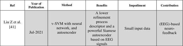
Tinnitus is a prevalent hearing disorder that can be caused by various factors such as age, hearing loss, exposure to loud noises, ear infections or tumors, certain medications, head or neck injuries, and psychological conditions like anxiety and depression. While not every patient requires medical attention, about 20% of sufferers seek clinical intervention. Early diagnosis is crucial for effective treatment. New developments have been made in tinnitus detection to aid in early detection of this illness. Over the past few years, there has been a notable growth in the usage of electroencephalography (EEG) to study variations in oscillatory brain activity related to tinnitus. However, the results obtained from numerous studies vary greatly, leading to conflicting conclusions. Currently, clinicians rely solely on their expertise to identify individuals with tinnitus. Researchers in this field have incorporated various data modalities and machine-learning techniques to aid clinicians in identifying tinnitus characteristics and classifying people with tinnitus. The purpose of writing this article is to review articles that focus on using machine learning (ML) to identify or predict tinnitus patients using EEG signals as input data. We have evaluated 11 articles published between 2016 and 2023 using a systematic literature review (SLR) method. This article arranges perfect summaries of all the research reviewed and compares the significant aspects of each. Additionally, we performed statistical analyses to gain a deeper comprehension of the most recent research in this area. Almost all of the reviewed articles followed a five-step procedure to achieve the goal of tinnitus. Disclosure. Finally, we discuss the open affairs and challenges in this method of tinnitus recognition or prediction and suggest future directions for research.
Alzheimers Disease Diagnosis by Deep Learning Using MRI-Based Approaches
Oct 26, 2023The most frequent kind of dementia of the nervous system, Alzheimer's disease, weakens several brain processes (such as memory) and eventually results in death. The clinical study uses magnetic resonance imaging to diagnose AD. Deep learning algorithms are capable of pattern recognition and feature extraction from the inputted raw data. As early diagnosis and stage detection are the most crucial elements in enhancing patient care and treatment outcomes, deep learning algorithms for MRI images have recently allowed for diagnosing a medical condition at the beginning stage and identifying particular symptoms of Alzheimer's disease. As a result, we aimed to analyze five specific studies focused on AD diagnosis using MRI-based deep learning algorithms between 2021 and 2023 in this study. To completely illustrate the differences between these techniques and comprehend how deep learning algorithms function, we attempted to explore selected approaches in depth.
Deep Learning Techniques for Cervical Cancer Diagnosis based on Pathology and Colposcopy Images
Oct 25, 2023Cervical cancer is a prevalent disease affecting millions of women worldwide every year. It requires significant attention, as early detection during the precancerous stage provides an opportunity for a cure. The screening and diagnosis of cervical cancer rely on cytology and colposcopy methods. Deep learning, a promising technology in computer vision, has emerged as a potential solution to improve the accuracy and efficiency of cervical cancer screening compared to traditional clinical inspection methods that are prone to human error. This review article discusses cervical cancer and its screening processes, followed by the Deep Learning training process and the classification, segmentation, and detection tasks for cervical cancer diagnosis. Additionally, we explored the most common public datasets used in both cytology and colposcopy and highlighted the popular and most utilized architectures that researchers have applied to both cytology and colposcopy. We reviewed 24 selected practical papers in this study and summarized them. This article highlights the remarkable efficiency in enhancing the precision and speed of cervical cancer analysis by Deep Learning, bringing us closer to early diagnosis and saving lives.
CRC-ICM: Colorectal Cancer Immune Cell Markers Pattern Dataset
Aug 19, 2023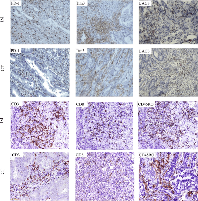
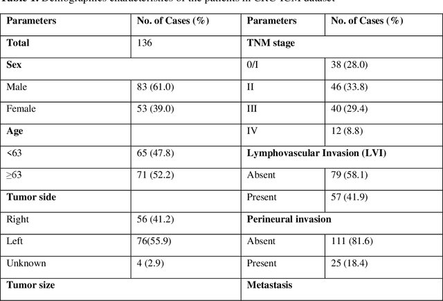
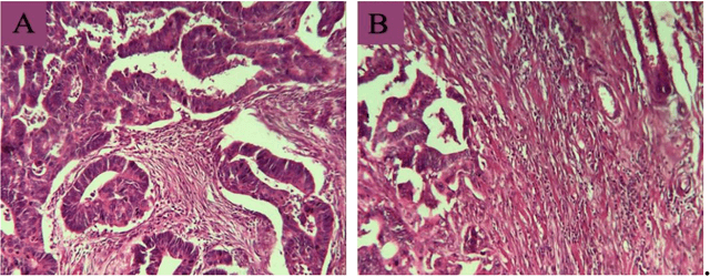

Colorectal Cancer (CRC) is the second most common cause of cancer death in the world, ad can be identified by the location of the primary tumor in the large intestine: right and left colon, and rectum. Based on the location, CRC shows differences in chromosomal and molecular characteristics, microbiomes incidence, pathogenesis, and outcome. It has been shown that tumors on left and right sides also have different immune landscape, so the prognosis may be different based on the primary tumor locations. It is widely accepted that immune components of the tumor microenvironment (TME) plays a critical role in tumor development. One of the critical regulatory molecules in the TME is immune checkpoints that as the gatekeepers of immune responses regulate the infiltrated immune cell functions. Inhibitory immune checkpoints such as PD-1, Tim3, and LAG3, as the main mechanism of immune suppression in TME overexpressed and result in further development of the tumor. The images of this dataset have been taken from colon tissues of patients with CRC, stained with specific antibodies for CD3, CD8, CD45RO, PD-1, LAG3 and Tim3. The name of this dataset is CRC-ICM and contains 1756 images related to 136 patients. The initial version of CRC-ICM is published on Elsevier Mendeley dataset portal, and the latest version is accessible via: https://databiox.com
Gastrointestinal Mucosal Problems Classification with Deep Learning
Jul 30, 2023Gastrointestinal mucosal changes can cause cancers after some years and early diagnosing them can be very useful to prevent cancers and early treatment. In this article, 8 classes of mucosal changes and anatomical landmarks including Polyps, Ulcerative Colitis, Esophagitis, Normal Z-Line, Normal Pylorus, Normal Cecum, Dyed Lifted Polyps, and Dyed Lifted Margin were predicted by deep learning. We used neural networks in this article. It is a black box artificial intelligence algorithm that works like a human neural system. In this article, Transfer Learning (TL) based on the Convolutional Neural Networks (CNNs), which is one of the well-known types of neural networks in image processing is used. We compared some famous CNN architecture including VGG, Inception, Xception, and ResNet. Our best model got 93% accuracy in test images. At last, we used our model in some real endoscopy and colonoscopy movies to classify problems.
ERCPMP: An Endoscopic Image and Video Dataset for Colorectal Polyps Morphology and Pathology
Jul 28, 2023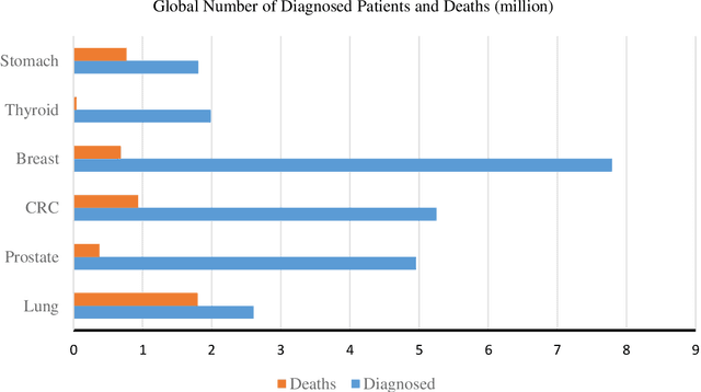
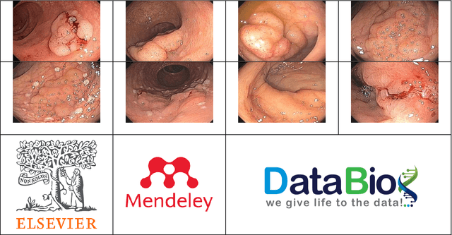
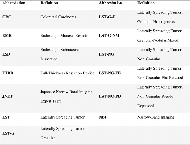
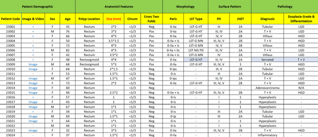
In the recent years, artificial intelligence (AI) and its leading subtypes, machine learning (ML) and deep learning (DL) and their applications are spreading very fast in various aspects such as medicine. Today the most important challenge of developing accurate algorithms for medical prediction, detection, diagnosis, treatment and prognosis is data. ERCPMP is an Endoscopic Image and Video Dataset for Recognition of Colorectal Polyps Morphology and Pathology. This dataset contains demographic, morphological and pathological data, endoscopic images and videos of 191 patients with colorectal polyps. Morphological data is included based on the latest international gastroenterology classification references such as Paris, Pit and JNET classification. Pathological data includes the diagnosis of the polyps including Tubular, Villous, Tubulovillous, Hyperplastic, Serrated, Inflammatory and Adenocarcinoma with Dysplasia Grade & Differentiation. The current version of this dataset is published and available on Elsevier Mendeley Dataverse and since it is under development, the latest version is accessible via: https://databiox.com.
A Survey on the Role of Artificial Intelligence in the Prediction and Diagnosis of Schizophrenia
May 19, 2023Machine learning is employed in healthcare to draw approximate conclusions regarding human diseases and mental health problems. Compared to older traditional methods, it can help to analyze data more efficiently and produce better and more dependable results. Millions of people are affected by schizophrenia, which is a chronic mental disorder that can significantly impact their lives. Many machine learning algorithms have been developed to predict and prevent this disease, and they can potentially be implemented in the diagnosis of individuals who have it. This survey aims to review papers that have focused on the use of deep learning to detect and predict schizophrenia using EEG signals, functional magnetic resonance imaging (fMRI), and diffusion magnetic resonance imaging (dMRI). With our chosen search strategy, we assessed ten publications from 2019 to 2022. All studies achieved successful predictions of more than 80%. This review provides summaries of the studies and compares their notable aspects. In the field of artificial intelligence (AI) and machine learning (ML) for schizophrenia, significant advances have been made due to the availability of ML tools, and we are optimistic that this field will continue to grow.