 Add to Chrome
 Add to Firefox
 Add to Edge
Medico Multimedia Task at MediaEval 2020: Automatic Polyp Segmentation
Dec 30, 2020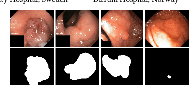

Colorectal cancer is the third most common cause of cancer worldwide. According to Global cancer statistics 2018, the incidence of colorectal cancer is increasing in both developing and developed countries. Early detection of colon anomalies such as polyps is important for cancer prevention, and automatic polyp segmentation can play a crucial role for this. Regardless of the recent advancement in early detection and treatment options, the estimated polyp miss rate is still around 20\%. Support via an automated computer-aided diagnosis system could be one of the potential solutions for the overlooked polyps. Such detection systems can help low-cost design solutions and save doctors time, which they could for example use to perform more patient examinations. In this paper, we introduce the 2020 Medico challenge, provide some information on related work and the dataset, describe the task and evaluation metrics, and discuss the necessity of organizing the Medico challenge.
DoubleU-Net: A Deep Convolutional Neural Network for Medical Image Segmentation
Jun 27, 2020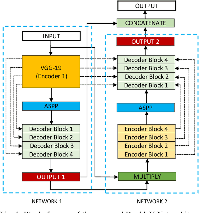
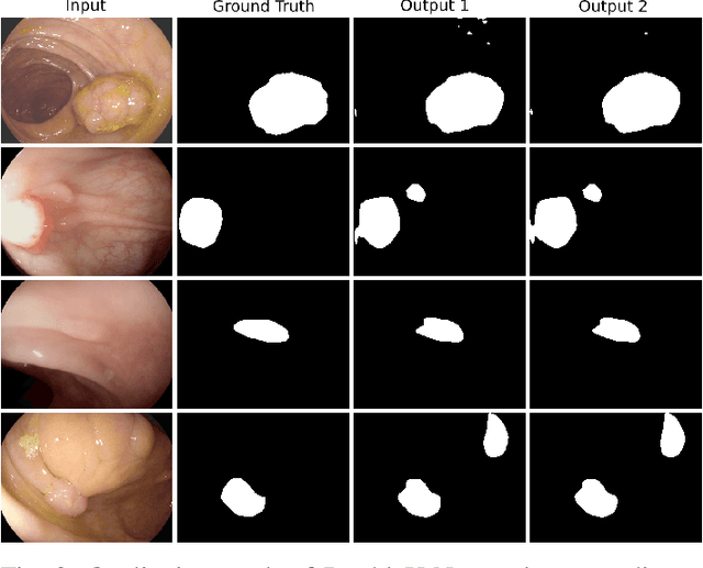
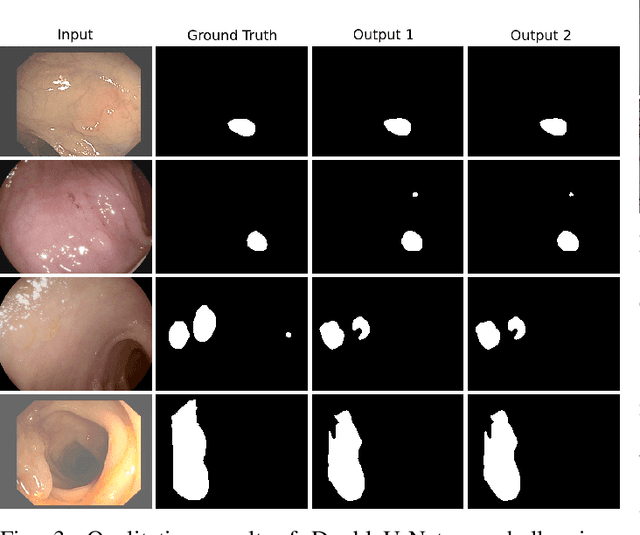
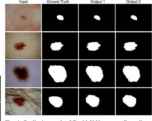
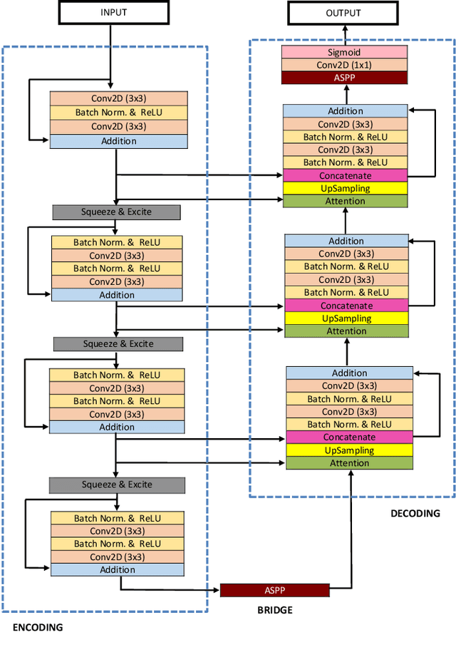
Semantic image segmentation is the process of labeling each pixel of an image with its corresponding class. An encoder-decoder based approach, like U-Net and its variants, is a popular strategy for solving medical image segmentation tasks. To improve the performance of U-Net on various segmentation tasks, we propose a novel architecture called DoubleU-Net, which is a combination of two U-Net architectures stacked on top of each other. The first U-Net uses a pre-trained VGG-19 as the encoder, which has already learned features from ImageNet and can be transferred to another task easily. To capture more semantic information efficiently, we added another U-Net at the bottom. We also adopt Atrous Spatial Pyramid Pooling (ASPP) to capture contextual information within the network. We have evaluated DoubleU-Net using four medical segmentation datasets, covering various imaging modalities such as colonoscopy, dermoscopy, and microscopy. Experiments on the MICCAI 2015 segmentation challenge, the CVC-ClinicDB, the 2018 Data Science Bowl challenge, and the Lesion boundary segmentation datasets demonstrate that the DoubleU-Net outperforms U-Net and the baseline models. Moreover, DoubleU-Net produces more accurate segmentation masks, especially in the case of the CVC-ClinicDB and MICCAI 2015 segmentation challenge datasets, which have challenging images such as smaller and flat polyps. These results show the improvement over the existing U-Net model. The encouraging results, produced on various medical image segmentation datasets, show that DoubleU-Net can be used as a strong baseline for both medical image segmentation and cross-dataset evaluation testing to measure the generalizability of Deep Learning (DL) models.
An Extensive Study on Cross-Dataset Bias and Evaluation Metrics Interpretation for Machine Learning applied to Gastrointestinal Tract Abnormality Classification
May 08, 2020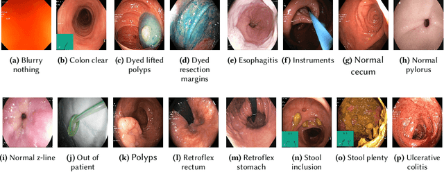
Precise and efficient automated identification of Gastrointestinal (GI) tract diseases can help doctors treat more patients and improve the rate of disease detection and identification. Currently, automatic analysis of diseases in the GI tract is a hot topic in both computer science and medical-related journals. Nevertheless, the evaluation of such an automatic analysis is often incomplete or simply wrong. Algorithms are often only tested on small and biased datasets, and cross-dataset evaluations are rarely performed. A clear understanding of evaluation metrics and machine learning models with cross datasets is crucial to bring research in the field to a new quality level. Towards this goal, we present comprehensive evaluations of five distinct machine learning models using Global Features and Deep Neural Networks that can classify 16 different key types of GI tract conditions, including pathological findings, anatomical landmarks, polyp removal conditions, and normal findings from images captured by common GI tract examination instruments. In our evaluation, we introduce performance hexagons using six performance metrics such as recall, precision, specificity, accuracy, F1-score, and Matthews Correlation Coefficient to demonstrate how to determine the real capabilities of models rather than evaluating them shallowly. Furthermore, we perform cross-dataset evaluations using different datasets for training and testing. With these cross-dataset evaluations, we demonstrate the challenge of actually building a generalizable model that could be used across different hospitals. Our experiments clearly show that more sophisticated performance metrics and evaluation methods need to be applied to get reliable models rather than depending on evaluations of the splits of the same dataset, i.e., the performance metrics should always be interpreted together rather than relying on a single metric.
Kvasir-SEG: A Segmented Polyp Dataset
Nov 16, 2019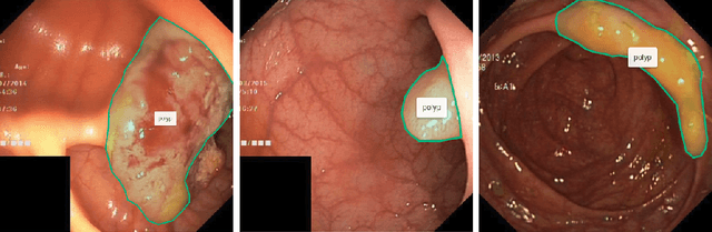
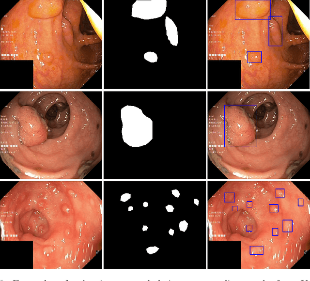
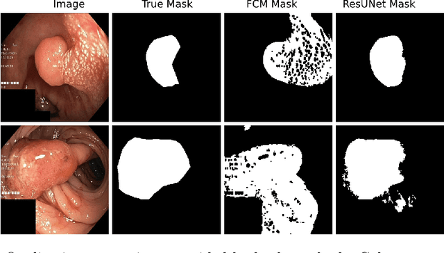
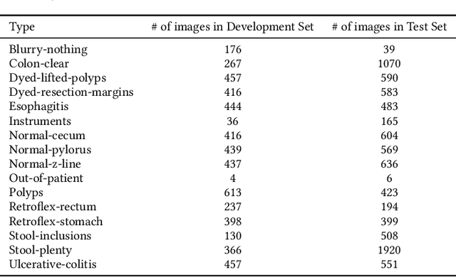
Pixel-wise image segmentation is a highly demanding task in medical-image analysis. In practice, it is difficult to find annotated medical images with corresponding segmentation masks. In this paper, we present Kvasir-SEG: an open-access dataset of gastrointestinal polyp images and corresponding segmentation masks, manually annotated by a medical doctor and then verified by an experienced gastroenterologist. Moreover, we also generated the bounding boxes of the polyp regions with the help of segmentation masks. We demonstrate the use of our dataset with a traditional segmentation approach and a modern deep-learning based Convolutional Neural Network (CNN) approach. The dataset will be of value for researchers to reproduce results and compare methods. By adding segmentation masks to the Kvasir dataset, which only provide frame-wise annotations, we enable multimedia and computer vision researchers to contribute in the field of polyp segmentation and automatic analysis of colonoscopy images.
ResUNet++: An Advanced Architecture for Medical Image Segmentation
Nov 16, 2019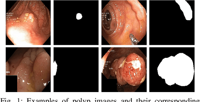
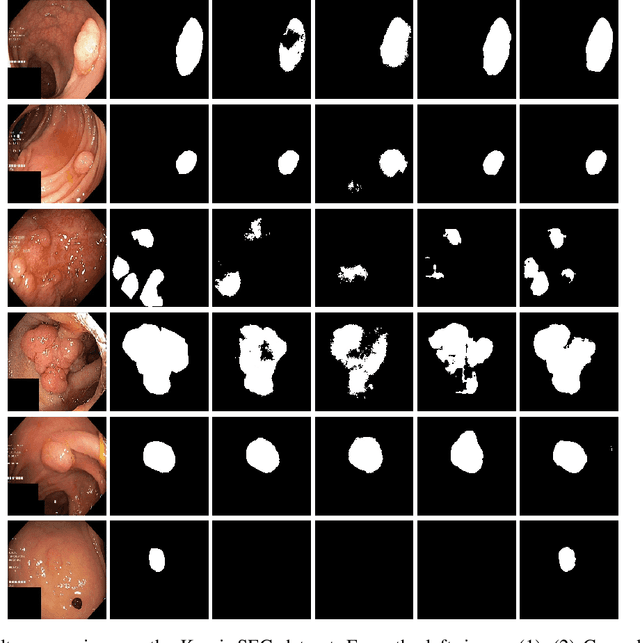
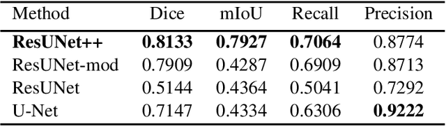
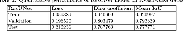
Accurate computer-aided polyp detection and segmentation during colonoscopy examinations can help endoscopists resect abnormal tissue and thereby decrease chances of polyps growing into cancer. Towards developing a fully automated model for pixel-wise polyp segmentation, we propose ResUNet++, which is an improved ResUNet architecture for colonoscopic image segmentation. Our experimental evaluations show that the suggested architecture produces good segmentation results on publicly available datasets. Furthermore, ResUNet++ significantly outperforms U-Net and ResUNet, two key state-of-the-art deep learning architectures, by achieving high evaluation scores with a dice coefficient of 81.33%, and a mean Intersection over Union (mIoU) of 79.27% for the Kvasir-SEG dataset and a dice coefficient of 79.55%, and a mIoU of 79.62% with CVC-612 dataset.
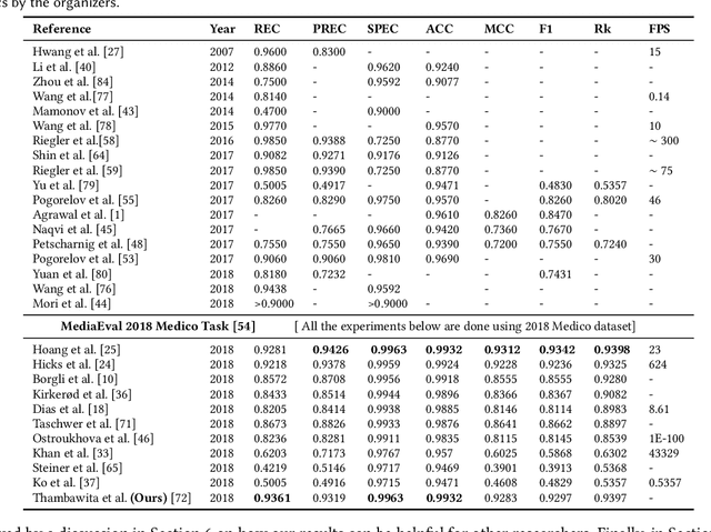
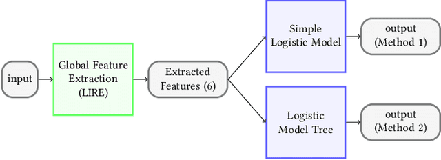
The Medico-Task 2018: Disease Detection in the Gastrointestinal Tract using Global Features and Deep Learning
Oct 31, 2018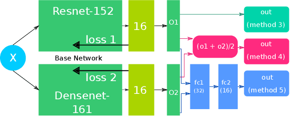
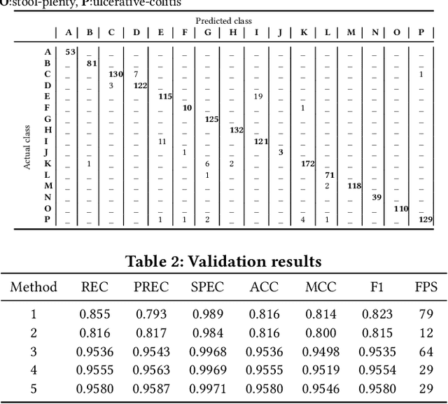
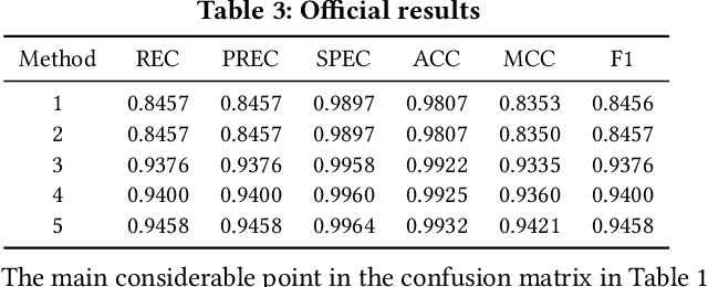
In this paper, we present our approach for the 2018 Medico Task classifying diseases in the gastrointestinal tract. We have proposed a system based on global features and deep neural networks. The best approach combines two neural networks, and the reproducible experimental results signify the efficiency of the proposed model with an accuracy rate of 95.80%, a precision of 95.87%, and an F1-score of 95.80%.
* 2 pages + 1 page for references, 1 figure, Conference paper