Effective and non-invasive radiological imaging based tumor/lesion characterization (e.g., subtype classification) has long been a major aim in the oncology diagnosis and treatment procedures, with the hope of reducing needs for invasive surgical biopsies. Prior work are generally very restricted to a limited patient sample size, especially using patient studies with confirmed pathological reports as ground truth. In this work, we curate a patient cohort of 1305 dynamic contrast CT studies (i.e., 5220 multi-phase 3D volumes) with pathology confirmed ground truth. A novel fully-automated and multi-stage liver tumor characterization framework is proposed, comprising four steps of tumor proposal detection, tumor harvesting, primary tumor site selection, and deep texture-based characterization. More specifically, (1) we propose a 3D non-isotropic anchor-free lesion detection method; (2) we present and validate the use of multi-phase deep texture learning for precise liver lesion tissue characterization, named spatially adaptive deep texture (SaDT); (3) we leverage small-sized public datasets to semi-automatically curate our large-scale clinical dataset of 1305 patients where four main liver tumor subtypes of primary, secondary, metastasized and benign are presented. Extensive evaluations demonstrate that our new data curation strategy, combined with the SaDT deep dynamic texture analysis, can effectively improve the mean F1 scores by >8.6% compared with baselines, in differentiating four major liver lesion types. This is a significant step towards the clinical goal.
As the demand for more descriptive machine learning models grows within medical imaging, bottlenecks due to data paucity will exacerbate. Thus, collecting enough large-scale data will require automated tools to harvest data/label pairs from messy and real-world datasets, such as hospital PACS. This is the focus of our work, where we present a principled data curation tool to extract multi-phase CT liver studies and identify each scan's phase from a real-world and heterogenous hospital PACS dataset. Emulating a typical deployment scenario, we first obtain a set of noisy labels from our institutional partners that are text mined using simple rules from DICOM tags. We train a deep learning system, using a customized and streamlined 3D SE architecture, to identify non-contrast, arterial, venous, and delay phase dynamic CT liver scans, filtering out anything else, including other types of liver contrast studies. To exploit as much training data as possible, we also introduce an aggregated cross entropy loss that can learn from scans only identified as "contrast". Extensive experiments on a dataset of 43K scans of 7680 patient imaging studies demonstrate that our 3DSE architecture, armed with our aggregated loss, can achieve a mean F1 of 0.977 and can correctly harvest up to 92.7% of studies, which significantly outperforms the text-mined and standard-loss approach, and also outperforms other, and more complex, model architectures.
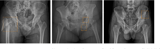
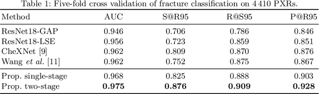
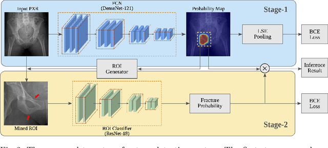
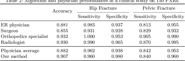
Hip and pelvic fractures are serious injuries with life-threatening complications. However, diagnostic errors of fractures in pelvic X-rays (PXRs) are very common, driving the demand for computer-aided diagnosis (CAD) solutions. A major challenge lies in the fact that fractures are localized patterns that require localized analyses. Unfortunately, the PXRs residing in hospital picture archiving and communication system do not typically specify region of interests. In this paper, we propose a two-stage hip and pelvic fracture detection method that executes localized fracture classification using weakly supervised ROI mining. The first stage uses a large capacity fully-convolutional network, i.e., deep with high levels of abstraction, in a multiple instance learning setting to automatically mine probable true positive and definite hard negative ROIs from the whole PXR in the training data. The second stage trains a smaller capacity model, i.e., shallower and more generalizable, with the mined ROIs to perform localized analyses to classify fractures. During inference, our method detects hip and pelvic fractures in one pass by chaining the probability outputs of the two stages together. We evaluate our method on 4 410 PXRs, reporting an area under the ROC curve value of 0.975, the highest among state-of-the-art fracture detection methods. Moreover, we show that our two-stage approach can perform comparably to human physicians (even outperforming emergency physicians and surgeons), in a preliminary reader study of 23 readers.