 Add to Chrome
 Add to Firefox
 Add to Edge
Synthesising clinically realistic Chest X-rays using Generative Adversarial Networks
Oct 07, 2020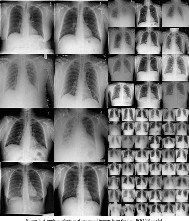
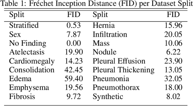
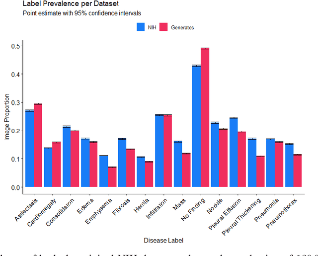
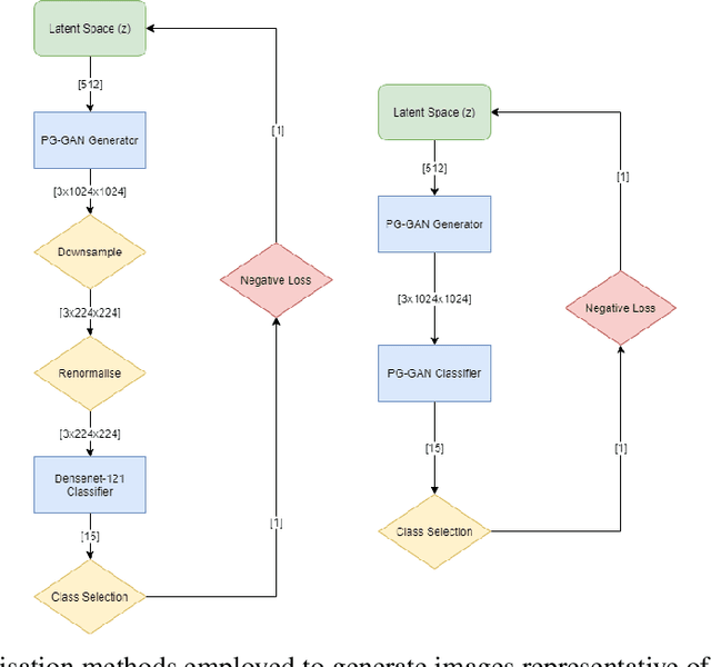
Chest x-rays are one of the most commonly performed medical investigations globally and are vital to identifying a number of conditions. These images are however protected under patient confidentiality and as such require the removal of identifying information as well as ethical clearance to be released. Generative adversarial networks (GANs) are a branch of deep learning which are capable of producing synthetic samples of a desired distribution. Image generation is one such application with recent advances enabling the production of high-resolution images, a feature vital to the utility of x-rays given the scale of various pathologies. We apply the Progressive Growing GAN (PGGAN) to the task of chest x-ray generation with the goal of being able to produce images without any ethical concerns that may be used for medical education or in other machine learning work. We evaluate the properties of the generated x-rays with a practicing radiologist and demonstrate that high-quality, realistic images can be produced with global features consistent with pathologies seen in the NIH dataset. Improvements in the reproduction of small-scale details remains for future work. We train a classification model on the NIH images and evaluate the distribution of disease labels across the generated samples. We find that the model is capable of reproducing all the abnormalities in a similar proportion to the source image distribution as labelled by the classifier. We additionally demonstrate that the latent space can be optimised to produce images of a particular class despite unconditional training, with the model producing related features and complications for the class of interest. We also validate the application of the Fr'echet Inception Distance (FID) to x-ray images and determine that the PGGAN reproduces x-ray images with an FID of 8.02, which is similar to other high resolution tasks.
Estimation of Body Mass Index from Photographs using Deep Convolutional Neural Networks
Aug 29, 2019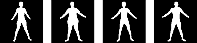

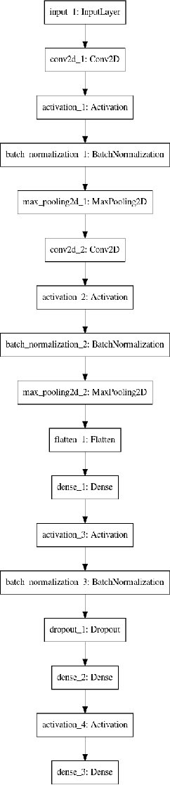
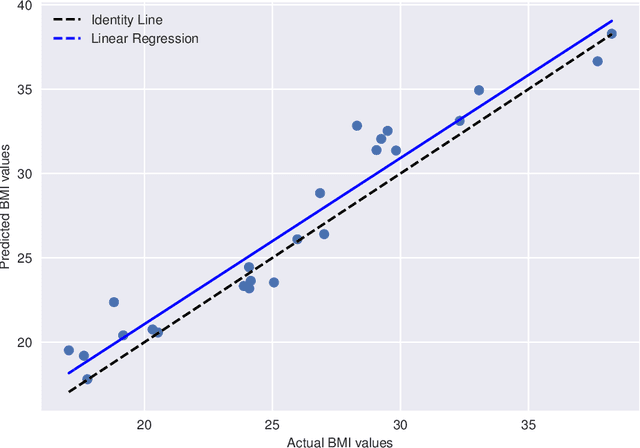
Obesity is an important concern in public health, and Body Mass Index is one of the useful (and proliferant) measures. We use Convolutional Neural Networks to determine Body Mass Index from photographs in a study with 161 participants. Low data, a common problem in medicine, is addressed by reducing the information in the photographs by generating silhouette images. Results present with high correlation when tested on unseen data.
Robotic Arm for Remote Surgery
Jul 22, 2013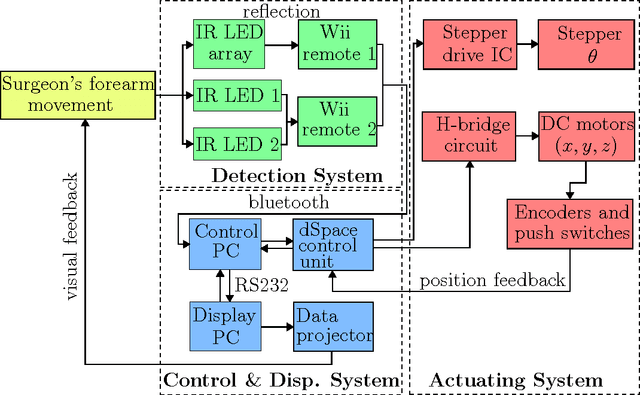
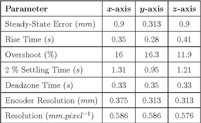
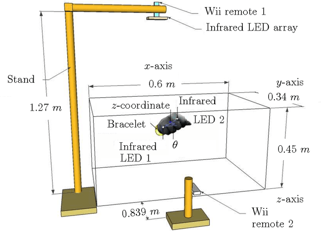
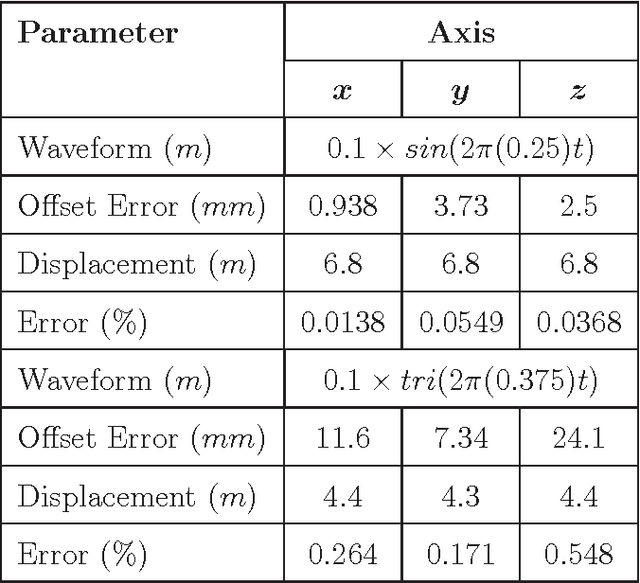
Recent advances in telecommunications have enabled surgeons to operate remotely on patients with the use of robotics. The investigation and testing of remote surgery using a robotic arm is presented. The robotic arm is designed to have four degrees of freedom that track the surgeon's x, y, z positions and the rotation angle of the forearm {\theta}. The system comprises two main subsystems viz. the detecting and actuating systems. The detection system uses infrared light-emitting diodes, a retroreflective bracelet and two infrared cameras which as a whole determine the coordinates of the surgeon's forearm. The actuation system, or robotic arm, is based on a lead screw mechanism which can obtain a maximum speed of 0.28 m/s with a 1.5 degree/step for the end-effector. The infrared detection and encoder resolutions are below 0.6 mm/pixel and 0.4 mm respectively, which ensures the robotic arm can operate precisely. The surgeon is able to monitor the patient with the use of a graphical user interface on the display computer. The lead screw system is modelled and compared to experimentation results. The system is controlled using a simple proportional-integrator (PI) control scheme which is implemented on a dSpace control unit. The control design results in a rise time of less than 0.5 s, a steady-state error of less than 1 mm and settling time of less than 1.4 s. The system accumulates, over an extended period of time, an error of approximately 4 mm due to inertial effects of the robotic arm. The results show promising system performance characteristics for a relatively inexpensive solution to a relatively advanced application.