 Add to Chrome
 Add to Firefox
 Add to Edge
An Approach for Thyroid Nodule Analysis Using Thermographic Images
May 28, 2026
Thyroid cancer is said to be the second most common type of cancer in female individuals and the third in males by 2030, according to projections. In general, detecting cancer in its early stages improves the chance of survival of the individual. Thermography is a diagnostic tool that has been increasingly used to detect cancer and abnormalities, including that of thyroid. Various methods to segment and detect hot regions in thermograms and, consequently, to detect suspicious tissues present in these images have been proposed. It is well known that medical diagnosis yields a great deal of information. Thus, physicians have to comprehensively analyse and evaluate this information in a short period of time, which is infeasible in most cases. In this work, we perform a general review of thermography , focusing on the thyroid analysis. We propose protocols for image acquisiton and an autonomous registration for thyroid images. We also perform analyses of the image data, which include feature extraction, image processing, and a possible approach for classification of healthy or unhealthy patients. In summary, this work presents a pilot project for detection of tumors in our university hospital, which is part of an effort to support preventive medical actions in our endocrinology department. Under some future adjustments, this project will be submitted for approval by the ethics and research committee of Hospital Universitário Antonio Pedro at Universidade Federal Fluminense (HUAP-UFF) and to the Brazilian Ministry of Health Ethical committee under the name: Evaluation of the importance of thermography to aid diagnosis of thyroid nodules of patients in HUAP-UFF (in Portuguese: Avaliação da importância da termografia no auxílio à investigação diagnóstica de nódulos tireoidianos em pacientes acompanhados no HUAP-UFF).

Comparing Results of Thermographic Images Based Diagnosis for Breast Diseases
Aug 30, 2022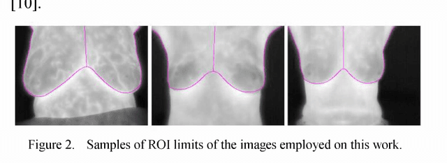
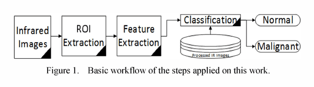
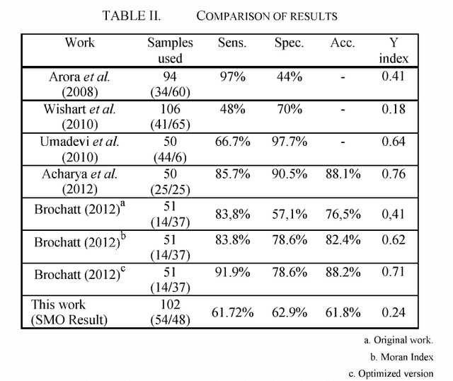
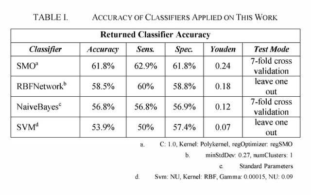
This paper examines the potential contribution of infrared (IR) imaging in breast diseases detection. It compares obtained results using some algorithms for detection of malignant breast conditions such as Support Vector Machine (SVM) regarding the consistency of different approaches when applied to public data. Moreover, in order to avail the actual IR imaging's capability as a complement on clinical trials and to promote researches using high-resolution IR imaging we deemed the use of a public database revised by confidently trained breast physicians as essential. Only the static acquisition protocol is regarded in our work. We used lO2 IR single breast images from the Pro Engenharia (PROENG) public database (54 normal and 48 with some finding). These images were collected from Universidade Federal de Pernambuco (UFPE) University's Hospital. We employed the same features proposed by the authors of the work that presented the best results and achieved an accuracy of 61.7 % and Youden index of 0.24 using the Sequential Minimal Optimization (SMO) classifier.