 Add to Chrome
 Add to Firefox
 Add to Edge
Dual Co-Train: Cross-Dataset Ultrasound Tongue Segmentation Under Extreme Data Scarcity
Aug 18, 2026
Ultrasound tongue contour segmentation remains challenging under cross-dataset domain shift, where limited annotations, probe variability, and acquisition noise often degrade model generalization. We present a source-free domain adaptation framework for robust ultrasound tongue segmentation built on a lightweight UltraUNet backbone. Starting from a checkpoint pretrained on only five labeled source images, simulating an underfitted constrained source model, the proposed method adapts to a fully-unlabeled target domain by iteratively refining pseudo-labels, filtering unreliable masks with a contour-based quality-control module, and generating target-style synthetic image-mask pairs through a segmentation-guided conditional GAN. The student model is then trained on a mixture of clean pseudo-labeled target images, noisy pseudo-labels with consistency regularization, and synthetic samples, enabling closed-loop adaptation without access to source data. We evaluate the method on 12 source-target transfer pairs across eight ultrasound tongue imaging datasets, and conduct source-size scaling experiments and ablation studies. Across all comparisons, the proposed framework improves segmentation overlap and contour accuracy over the baselines, including supervised ones. These results suggest that task-specific pseudo-label refinement and synthetic target-style augmentation can substantially improve source-free adaptation for ultrasound tongue imaging.

Morphological-consistent Diffusion Network for Ultrasound Coronal Image Enhancement
Sep 25, 2024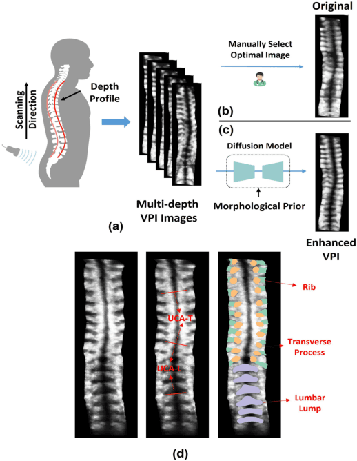
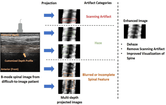
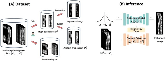
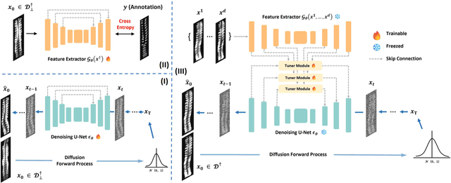
Ultrasound curve angle (UCA) measurement provides a radiation-free and reliable evaluation for scoliosis based on ultrasound imaging. However, degraded image quality, especially in difficult-to-image patients, can prevent clinical experts from making confident measurements, even leading to misdiagnosis. In this paper, we propose a multi-stage image enhancement framework that models high-quality image distribution via a diffusion-based model. Specifically, we integrate the underlying morphological information from images taken at different depths of the 3D volume to calibrate the reverse process toward high-quality and high-fidelity image generation. This is achieved through a fusion operation with a learnable tuner module that learns the multi-to-one mapping from multi-depth to high-quality images. Moreover, the separate learning of the high-quality image distribution and the spinal features guarantees the preservation of consistent spinal pose descriptions in the generated images, which is crucial in evaluating spinal deformities. Remarkably, our proposed enhancement algorithm significantly outperforms other enhancement-based methods on ultrasound images in terms of image quality. Ultimately, we conduct the intra-rater and inter-rater measurements of UCA and higher ICC (0.91 and 0.89 for thoracic and lumbar angles) on enhanced images, indicating our method facilitates the measurement of ultrasound curve angles and offers promising prospects for automated scoliosis diagnosis.
Automatic Ultrasound Curve Angle Measurement via Affinity Clustering for Adolescent Idiopathic Scoliosis Evaluation
May 07, 2024
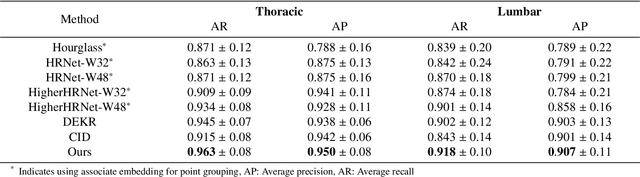
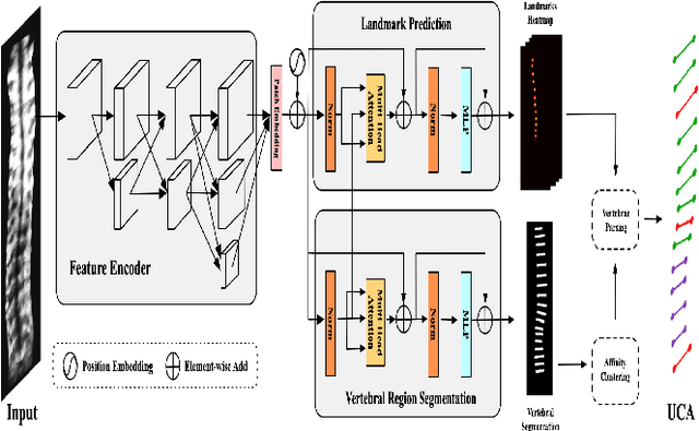
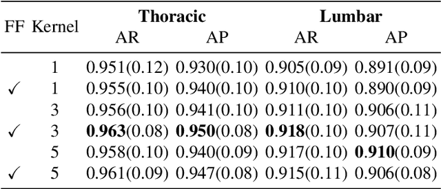
The current clinical gold standard for evaluating adolescent idiopathic scoliosis (AIS) is X-ray radiography, using Cobb angle measurement. However, the frequent monitoring of the AIS progression using X-rays poses a challenge due to the cumulative radiation exposure. Although 3D ultrasound has been validated as a reliable and radiation-free alternative for scoliosis assessment, the process of measuring spinal curvature is still carried out manually. Consequently, there is a considerable demand for a fully automatic system that can locate bony landmarks and perform angle measurements. To this end, we introduce an estimation model for automatic ultrasound curve angle (UCA) measurement. The model employs a dual-branch network to detect candidate landmarks and perform vertebra segmentation on ultrasound coronal images. An affinity clustering strategy is utilized within the vertebral segmentation area to illustrate the affinity relationship between candidate landmarks. Subsequently, we can efficiently perform line delineation from a clustered affinity map for UCA measurement. As our method is specifically designed for UCA calculation, this method outperforms other state-of-the-art methods for landmark and line detection tasks. The high correlation between the automatic UCA and Cobb angle (R$^2$=0.858) suggests that our proposed method can potentially replace manual UCA measurement in ultrasound scoliosis assessment.