 Add to Chrome
 Add to Firefox
 Add to Edge
Machine learning refinement of in situ images acquired by low electron dose LC-TEM
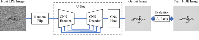
Oct 31, 2023We study a machine learning (ML) technique for refining images acquired during in situ observation using liquid-cell transmission electron microscopy (LC-TEM). Our model is constructed using a U-Net architecture and a ResNet encoder. For training our ML model, we prepared an original image dataset that contained pairs of images of samples acquired with and without a solution present. The former images were used as noisy images and the latter images were used as corresponding ground truth images. The number of pairs of image sets was $1,204$ and the image sets included images acquired at several different magnifications and electron doses. The trained model converted a noisy image into a clear image. The time necessary for the conversion was on the order of 10ms, and we applied the model to in situ observations using the software Gatan DigitalMicrograph (DM). Even if a nanoparticle was not visible in a view window in the DM software because of the low electron dose, it was visible in a successive refined image generated by our ML model.
Fast improvement of TEM image with low-dose electrons by deep learning
Jun 03, 2021
Low-electron-dose observation is indispensable for observing various samples using a transmission electron microscope; consequently, image processing has been used to improve transmission electron microscopy (TEM) images. To apply such image processing to in situ observations, we here apply a convolutional neural network to TEM imaging. Using a dataset that includes short-exposure images and long-exposure images, we develop a pipeline for processed short-exposure images, based on end-to-end training. The quality of images acquired with a total dose of approximately 5 e- per pixel becomes comparable to that of images acquired with a total dose of approximately 1000 e- per pixel. Because the conversion time is approximately 8 ms, in situ observation at 125 fps is possible. This imaging technique enables in situ observation of electron-beam-sensitive specimens.