 Add to Chrome
 Add to Firefox
 Add to Edge
CoM$^3$eT: A foundation model for medical image analysis through federated, multidimensional context integration
Aug 17, 2026
Medical foundation models improve generalization when training AI models with limited labeled data, but remain confined to a single specialty, such as pathology or radiology, and to either sparse or dense outputs, such as classification or segmentation. Here, we present CoM$^3$eT (Co-representation Multidimensional Multitask Medical Transformer), a medical vision foundation model that unifies pathology and radiology, sparse and dense predictions, and two- and higher-dimensional inputs by modeling multidimensional context with attention. CoM$^3$eT outperformed other medical foundation models in an open competition spanning five tomographic, four whole-specimen, and three two-dimensional datasets, covering sparse and dense prediction tasks as well as report generation. When adapted across diverse clinical applications, training fewer than 2.5% of parameters achieved performance comparable to full fine-tuning, enabling research without access to high-performance GPU clusters. Applied to federated learning across hospitals, this approach achieved performance comparable to pooled-data training over internet connections and with consumer-grade hardware.

Volumetric Reconstruction of Prostatectomy Specimens from Histology
Nov 29, 2024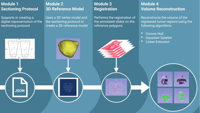
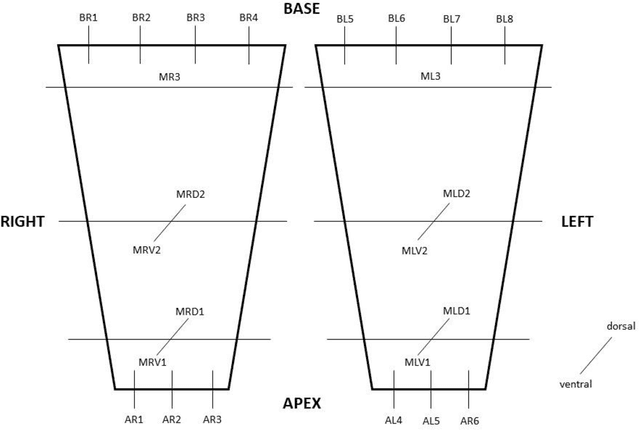
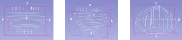
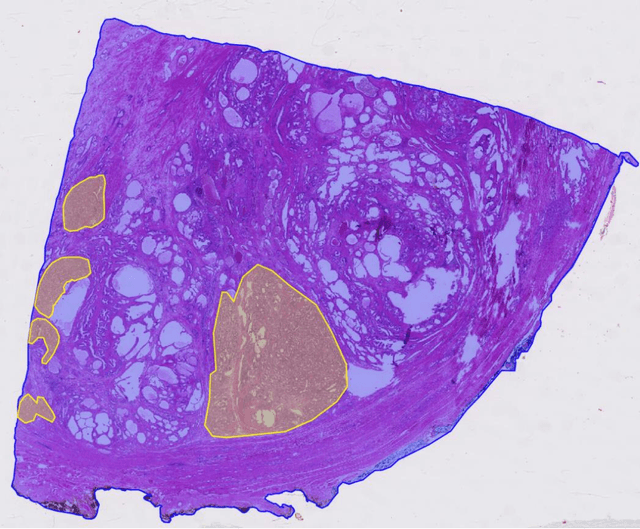
Surgical treatment for prostate cancer often involves organ removal, i.e., prostatectomy. Pathology reports on these specimens convey treatment-relevant information. Beyond these reports, the diagnostic process generates extensive and complex information that is difficult to represent in reports, although it is of significant interest to the other medical specialties involved. 3D tissue reconstruction would allow for better spatial visualization, as well as combinations with other imaging modalities. Existing approaches in this area have proven labor-intensive and challenging to integrate into clinical workflows. 3D-SLIVER provides a simplified solution, implemented as an open-source 3DSlicer extension. We outline three specific real-world scenarios to illustrate its potential to improve transparency in diagnostic workflows and contribute to multi-modal research endeavors. Implementing the 3D reconstruction process involved four sub-modules of 3D-SLIVER: digitization of slicing protocol, virtual slicing of arbitrary 3D models based on that protocol, registration of slides with virtual slices using the Coherent Point Drift algorithm, and 3D reconstruction of registered information using convex hulls, Gaussian splatter and linear extrusion. Three use cases to employ 3D-SLIVER are presented: a low-effort approach to pathology workflow integration and two research-related use cases illustrating how to perform retrospective evaluations of PI-RADS predictions and statistically model 3D distributions of morphological patterns. 3D-SLIVER allows for improved interdisciplinary communication among specialties. It is designed for simplicity in application, allowing for flexible integration into various workflows and use cases. Here we focused on the clinical care of prostate cancer patients, but future possibilities are extensive with other neoplasms and in education and research.