 Add to Chrome
 Add to Firefox
 Add to Edge
Designing UNICORN: a Unified Benchmark for Imaging in Computational Pathology, Radiology, and Natural Language
Mar 03, 2026
Medical foundation models show promise to learn broadly generalizable features from large, diverse datasets. This could be the base for reliable cross-modality generalization and rapid adaptation to new, task-specific goals, with only a few task-specific examples. Yet, evidence for this is limited by the lack of public, standardized, and reproducible evaluation frameworks, as existing public benchmarks are often fragmented across task-, organ-, or modality-specific settings, limiting assessment of cross-task generalization. We introduce UNICORN, a public benchmark designed to systematically evaluate medical foundation models under a unified protocol. To isolate representation quality, we built the benchmark on a novel two-step framework that decouples model inference from task-specific evaluation based on standardized few-shot adaptation. As a central design choice, we constructed indirectly accessible sequestered test sets derived from clinically relevant cohorts, along with standardized evaluation code and a submission interface on an open benchmarking platform. Performance is aggregated into a single UNICORN Score, a new metric that we introduce to support direct comparison of foundation models across diverse medical domains, modalities, and task types. The UNICORN test dataset includes data from more than 2,400 patients, including over 3,700 vision cases and over 2,400 clinical reports collected from 17 institutions across eight countries. The benchmark spans eight anatomical regions and four imaging modalities. Both task-specific and aggregated leaderboards enable accessible, standardized, and reproducible evaluation. By standardizing multi-task, multi-modality assessment, UNICORN establishes a foundation for reproducible benchmarking of medical foundation models. Data, baseline methods, and the evaluation platform are publicly available via unicorn.grand-challenge.org.

Stable prediction with radiomics data
Mar 27, 2019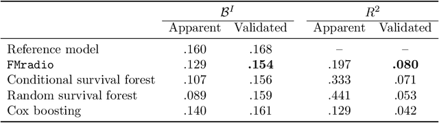
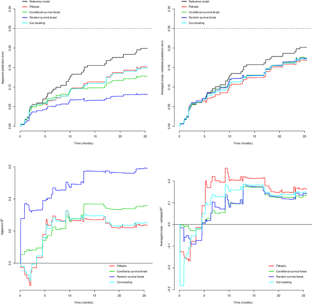
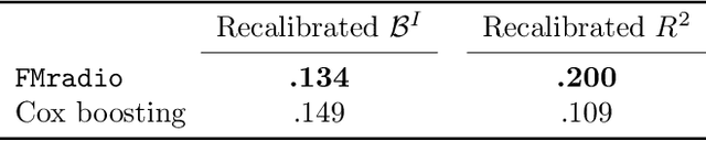
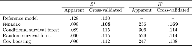
Motivation: Radiomics refers to the high-throughput mining of quantitative features from radiographic images. It is a promising field in that it may provide a non-invasive solution for screening and classification. Standard machine learning classification and feature selection techniques, however, tend to display inferior performance in terms of (the stability of) predictive performance. This is due to the heavy multicollinearity present in radiomic data. We set out to provide an easy-to-use approach that deals with this problem. Results: We developed a four-step approach that projects the original high-dimensional feature space onto a lower-dimensional latent-feature space, while retaining most of the covariation in the data. It consists of (i) penalized maximum likelihood estimation of a redundancy filtered correlation matrix. The resulting matrix (ii) is the input for a maximum likelihood factor analysis procedure. This two-stage maximum-likelihood approach can be used to (iii) produce a compact set of stable features that (iv) can be directly used in any (regression-based) classifier or predictor. It outperforms other classification (and feature selection) techniques in both external and internal validation settings regarding survival in squamous cell cancers.