 Add to Chrome
 Add to Firefox
 Add to Edge
Unsupervised Anomaly Detection for Image Dataset Quality Assurance in Multi-Center Breast MRI
Aug 17, 2026
Corrupted, inconsistent, or anomalous data silently threatens the safety and reliability of medical AI. Despite growing regulatory recognition of dataset quality assurance (QA) for high-risk medical AI, scalable automated detection remains underdeveloped. We employ unsupervised anomaly detection (AD) and out-of-distribution (OOD) detection as an automated dataset QA mechanism for multi-center dynamic contrast-enhanced breast MRI. We build a controlled AD benchmark of 17 realistic QA-relevant anomaly types from six public datasets (protocol violations, processing errors, incorrect anatomical regions) and propose a taxonomy of radiological image anomalies based on human visual perception, enabling fine-grained analysis of AD failure modes. The benchmark includes near-, medium-far-, far-OOD samples, as well as in-distribution and external normal data. Four methods are evaluated: a projection-based method extended with a domain-specific feature extractor and a novel positional encoding, a reconstruction-based approach extended to full 3D volumes with an augmented training objective, and two unmodified hybrid OOD detection methods. Medium-far- and far-OOD samples are detected reliably, whereas near-OOD samples and external normal data from unseen institutions expose method-specific differences. The 3D reconstruction-based approach best balances detection performance (AUROC: 0.936) and generalization to unseen institutions. The projection-based method with positional encoding achieves the highest overall detection performance (AUROC: 0.954). Both hybrid methods exhibit critical failure modes, confirming that methods validated for one modality or anatomy may not generalize without domain-specific adaptation. Implants and mastectomies remain an open challenge for all methods. Our results establish a foundation and practical guidance on scalable unsupervised QA in medical AI pipelines.

Tubular Shape Aware Data Generation for Semantic Segmentation in Medical Imaging
Oct 02, 2020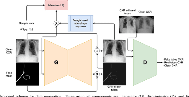
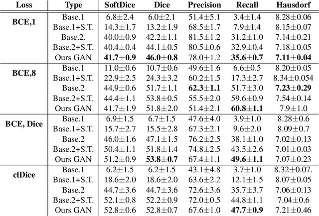
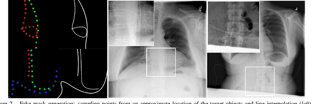
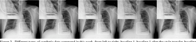
Chest X-ray is one of the most widespread examinations of the human body. In interventional radiology, its use is frequently associated with the need to visualize various tube-like objects, such as puncture needles, guiding sheaths, wires, and catheters. Detection and precise localization of these tube-like objects in the X-ray images is, therefore, of utmost value, catalyzing the development of accurate target-specific segmentation algorithms. Similar to the other medical imaging tasks, the manual pixel-wise annotation of the tubes is a resource-consuming process. In this work, we aim to alleviate the lack of the annotated images by using artificial data. Specifically, we present an approach for synthetic data generation of the tube-shaped objects, with a generative adversarial network being regularized with a prior-shape constraint. Our method eliminates the need for paired image--mask data and requires only a weakly-labeled dataset (10--20 images) to reach the accuracy of the fully-supervised models. We report the applicability of the approach for the task of segmenting tubes and catheters in the X-ray images, whereas the results should also hold for the other imaging modalities.