 Add to Chrome
 Add to Firefox
 Add to Edge
Automatic classification between COVID-19 pneumonia, non-COVID-19 pneumonia, and the healthy on chest X-ray image: combination of data augmentation methods
Jun 12, 2020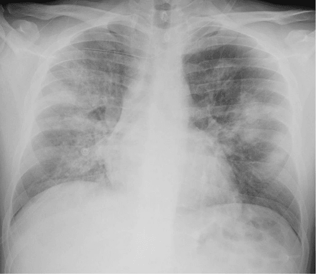
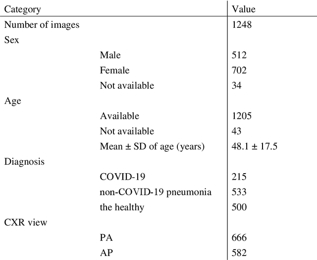
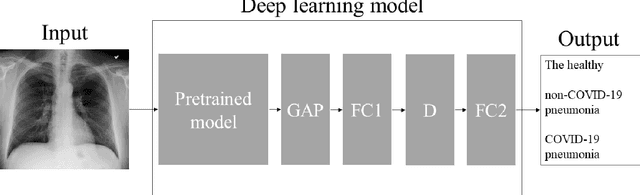
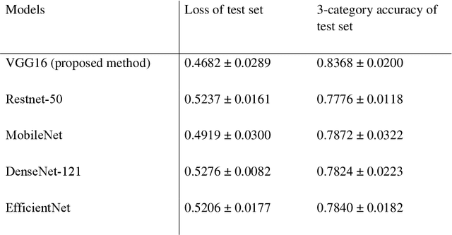
Purpose: This study aimed to develop and validate computer-aided diagnosis (CXDx) system for classification between COVID-19 pneumonia, non-COVID-19 pneumonia, and the healthy on chest X-ray (CXR) images. Materials and Methods: From two public datasets, 1248 CXR images were obtained, which included 215, 533, and 500 CXR images of COVID-19 pneumonia patients, non-COVID-19 pneumonia patients, and the healthy samples. The proposed CADx system utilized VGG16 as a pre-trained model and combination of conventional method and mixup as data augmentation methods. Other types of pre-trained models were compared with the VGG16-based model. Single type or no data augmentation methods were also evaluated. Splitting of training/validation/test sets was used when building and evaluating the CADx system. Three-category accuracy was evaluated for test set with 125 CXR images. Results: The three-category accuracy of the CAD system was 83.6% between COVID-19 pneumonia, non-COVID-19 pneumonia, and the healthy. Sensitivity for COVID-19 pneumonia was more than 90%. The combination of conventional method and mixup was more useful than single type or no data augmentation method. Conclusion: This study was able to create an accurate CADx system for the 3-category classification. Source code of our CADx system is available as open source for COVID-19 research.
Automatic detection of acute ischemic stroke using non-contrast computed tomography and two-stage deep learning model
Apr 09, 2020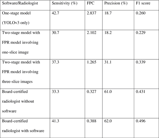
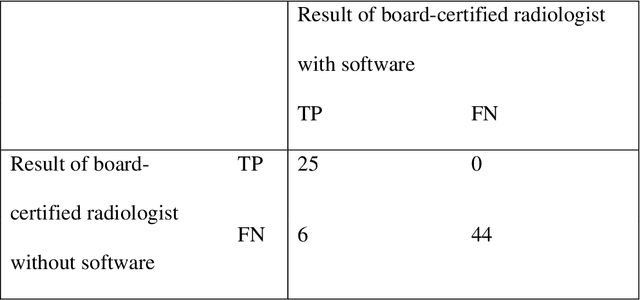
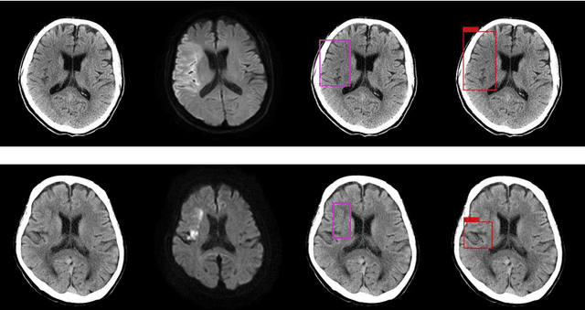
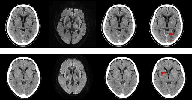
Background and Purpose: We aimed to develop and evaluate an automatic acute ischemic stroke-related (AIS) detection system involving a two-stage deep learning model. Methods: We included 238 cases from two different institutions. AIS-related findings were annotated on each of the 238 sets of head CT images by referring to head magnetic resonance imaging (MRI) images in which an MRI examination was performed within 24 h following the CT scan. These 238 annotated cases were divided into a training set including 189 cases and test set including 49 cases. Subsequently, a two-stage deep learning detection model was constructed from the training set using the You Only Look Once v3 model and Visual Geometry Group 16 classification model. Then, the two-stage model performed the AIS detection process in the test set. To assess the detection model's results, a board-certified radiologist also evaluated the test set head CT images with and without the aid of the detection model. The sensitivity of AIS detection and number of false positives were calculated for the evaluation of the test set detection results. The sensitivity of the radiologist with and without the software detection results was compared using the McNemar test. A p-value of less than 0.05 was considered statistically significant. Results: For the two-stage model and radiologist without and with the use of the software results, the sensitivity was 37.3%, 33.3%, and 41.3%, respectively, and the number of false positives per one case was 1.265, 0.327, and 0.388, respectively. On using the two-stage detection model's results, the board-certified radiologist's detection sensitivity significantly improved (p-value = 0.0313). Conclusions: Our detection system involving the two-stage deep learning model significantly improved the radiologist's sensitivity in AIS detection.