 Add to Chrome
 Add to Firefox
 Add to Edge
DALL-M: Context-Aware Clinical Data Augmentation with LLMs
Jul 11, 2024


X-ray images are vital in medical diagnostics, but their effectiveness is limited without clinical context. Radiologists often find chest X-rays insufficient for diagnosing underlying diseases, necessitating comprehensive clinical features and data integration. We present a novel technique to enhance the clinical context through augmentation techniques with clinical tabular data, thereby improving its applicability and reliability in AI medical diagnostics. To address this, we introduce a pioneering approach to clinical data augmentation that employs large language models (LLMs) to generate patient contextual synthetic data. This methodology is crucial for training more robust deep learning models in healthcare. It preserves the integrity of real patient data while enriching the dataset with contextually relevant synthetic features, significantly enhancing model performance. DALL-M uses a three-phase feature generation process: (i) clinical context storage, (ii) expert query generation, and (iii) context-aware feature augmentation. DALL-M generates new, clinically relevant features by synthesizing chest X-ray images and reports. Applied to 799 cases using nine features from the MIMIC-IV dataset, it created an augmented set of 91 features. This is the first work to generate contextual values for existing and new features based on patients' X-ray reports, gender, and age and to produce new contextual knowledge during data augmentation. Empirical validation with machine learning models, including Decision Trees, Random Forests, XGBoost, and TabNET, showed significant performance improvements. Incorporating augmented features increased the F1 score by 16.5% and Precision and Recall by approximately 25%. DALL-M addresses a critical gap in clinical data augmentation, offering a robust framework for generating contextually enriched datasets.
MDF-Net: Multimodal Dual-Fusion Network for Abnormality Detection using CXR Images and Clinical Data
Feb 26, 2023This study aims to investigate the effects of including patients' clinical information on the performance of deep learning (DL) classifiers for disease location in chest X-ray images. Although current classifiers achieve high performance using chest X-ray images alone, our interviews with radiologists indicate that clinical data is highly informative and essential for interpreting images and making proper diagnoses. In this work, we propose a novel architecture consisting of two fusion methods that enable the model to simultaneously process patients' clinical data (structured data) and chest X-rays (image data). Since these data modalities are in different dimensional spaces, we propose a spatial arrangement strategy, termed spatialization, to facilitate the multimodal learning process in a Mask R-CNN model. We performed an extensive experimental evaluation comprising three datasets with different modalities: MIMIC CXR (chest X-ray images), MIMIC IV-ED (patients' clinical data), and REFLACX (annotations of disease locations in chest X-rays). Results show that incorporating patients' clinical data in a DL model together with the proposed fusion methods improves the performance of disease localization in chest X-rays by 12\% in terms of Average Precision compared to a standard Mask R-CNN using only chest X-rays. Further ablation studies also emphasize the importance of multimodal DL architectures and the incorporation of patients' clinical data in disease localisation. The architecture proposed in this work is publicly available to promote the scientific reproducibility of our study (https://github.com/ChihchengHsieh/multimodal-abnormalities-detection).
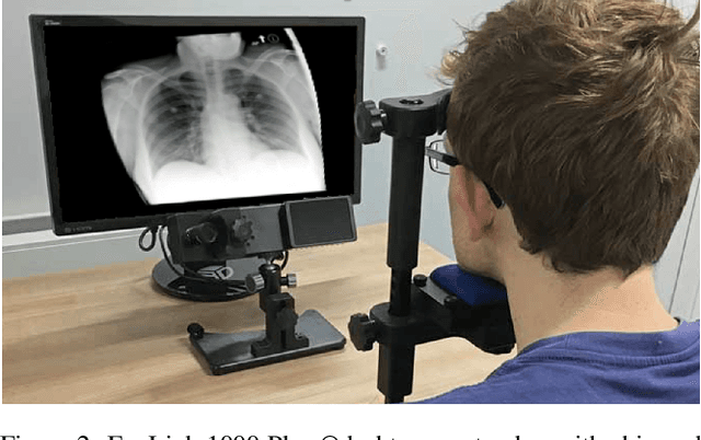
Integrating Eye-Gaze Data into CXR DL Approaches: A Preliminary study
Feb 06, 2023


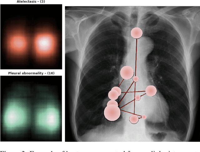
This paper proposes a novel multimodal DL architecture incorporating medical images and eye-tracking data for abnormality detection in chest x-rays. Our results show that applying eye gaze data directly into DL architectures does not show superior predictive performance in abnormality detection chest X-rays. These results support other works in the literature and suggest that human-generated data, such as eye gaze, needs a more thorough investigation before being applied to DL architectures.
Improving X-ray Diagnostics through Eye-Tracking and XR
Mar 03, 2022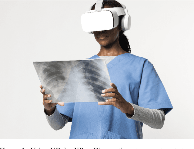
There is a growing need to assist radiologists in performing X-ray readings and diagnoses fast, comfortably, and effectively. As radiologists strive to maximize productivity, it is essential to consider the impact of reading rooms in interpreting complex examinations and ensure that higher volume and reporting speeds do not compromise patient outcomes. Virtual Reality (VR) is a disruptive technology for clinical practice in assessing X-ray images. We argue that conjugating eye-tracking with VR devices and Machine Learning may overcome obstacles posed by inadequate ergonomic postures and poor room conditions that often cause erroneous diagnostics when professionals examine digital images.