 Add to Chrome
 Add to Firefox
 Add to Edge
Does FLAIR super-resolution erase or hallucinate small white-matter lesions?
Aug 06, 2026
White matter hyperintensities (WMH), bright regions on Fluid-attenuated Inversion Recovery (FLAIR) scans are associated with cerebrovascular pathology and neurodegeneration. FLAIR is usually acquired with thick slices in clinical settings, giving it poor through-plane resolution. Super-resolution (SR) is a widely used method for recovering an isotropic volume from an anisotropic scan. Yet whether applying it prior to WMH segmentation preserves lesion content remains unknown: a model may erase small real lesions or hallucinate absent ones. We used 1-mm isotropic high-resolution (HR) FLAIR scans from 29 individuals in the ADNI cohort, each manually segmented for WMH by an expert. Then, we degraded each to simulated 3 and 5 mm through-plane acquisitions. Multi-contrast implicit neural representation (INR), a single-contrast self-supervised model (ECLARE), and cubic interpolation were used to upsample them onto the HR grid. WMH segmentation from a simulated thick slice and the original HR FLAIR set the floor and ceiling, respectively, for the per-lesion analysis. Of four WMH segmentation methods (WMH-SynthSeg, segcsvd, MARS-WMH, TrUE-Net), we ran the analysis under the most sensitive one to small lesions on HR (MARS-WMH) with the evaluation metrics of detection sensitivity, erasure rate (HR-detected lesions lost after reconstruction), and hallucination rate (predicted components absent from both the manual and HR segmentation). The dominant effect of SR was erasure of small real lesions, not hallucination, and it increased with slice thickness, though every reconstruction still improved lesion detection over the raw thick slice. ECLARE recovered small lesion signal best at both thicknesses, while the INR was no better than cubic interpolation.

Surface-based parcellation and vertex-wise analysis of ultra high-resolution ex vivo 7 tesla MRI in neurodegenerative diseases
Mar 28, 2024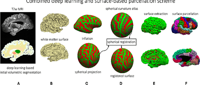
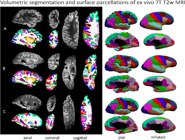
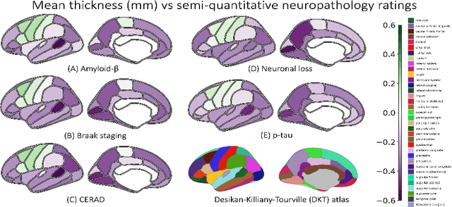
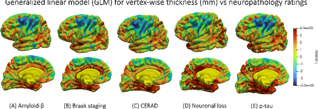
Magnetic resonance imaging (MRI) is the standard modality to understand human brain structure and function in vivo (antemortem). Decades of research in human neuroimaging has led to the widespread development of methods and tools to provide automated volume-based segmentations and surface-based parcellations which help localize brain functions to specialized anatomical regions. Recently ex vivo (postmortem) imaging of the brain has opened-up avenues to study brain structure at sub-millimeter ultra high-resolution revealing details not possible to observe with in vivo MRI. Unfortunately, there has been limited methodological development in ex vivo MRI primarily due to lack of datasets and limited centers with such imaging resources. Therefore, in this work, we present one-of-its-kind dataset of 82 ex vivo T2w whole brain hemispheres MRI at 0.3 mm isotropic resolution spanning Alzheimer's disease and related dementias. We adapted and developed a fast and easy-to-use automated surface-based pipeline to parcellate, for the first time, ultra high-resolution ex vivo brain tissue at the native subject space resolution using the Desikan-Killiany-Tourville (DKT) brain atlas. This allows us to perform vertex-wise analysis in the template space and thereby link morphometry measures with pathology measurements derived from histology. We will open-source our dataset docker container, Jupyter notebooks for ready-to-use out-of-the-box set of tools and command line options to advance ex vivo MRI clinical brain imaging research on the project webpage.
Improved Segmentation of Deep Sulci in Cortical Gray Matter Using a Deep Learning Framework Incorporating Laplace's Equation
Mar 03, 2023

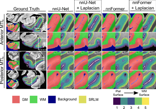

When developing tools for automated cortical segmentation, the ability to produce topologically correct segmentations is important in order to compute geometrically valid morphometry measures. In practice, accurate cortical segmentation is challenged by image artifacts and the highly convoluted anatomy of the cortex itself. To address this, we propose a novel deep learning-based cortical segmentation method in which prior knowledge about the geometry of the cortex is incorporated into the network during the training process. We design a loss function which uses the theory of Laplace's equation applied to the cortex to locally penalize unresolved boundaries between tightly folded sulci. Using an ex vivo MRI dataset of human medial temporal lobe specimens, we demonstrate that our approach outperforms baseline segmentation networks, both quantitatively and qualitatively.