 Add to Chrome
 Add to Firefox
 Add to Edge
The autoPET3 Challenge -- Automated Lesion Segmentation in Whole-Body PET/CT - Multitracer Multicenter Generalization
May 07, 2026We report the design and results of the third autoPET challenge (MICCAI 2024), which benchmarked automated lesion segmentation in whole-body PET/CT under a compositional generalization setting. Training data comprised 1,014 [18F]-FDG PET/CT studies from the University Hospital Tübingen and 597 [18F]/[68Ga]-PSMA PET/CT studies from the LMU University Hospital Munich, constituting the largest publicly available annotated PSMA PET/CT dataset to date. The held-out test set of 200 studies covered four tracer-center combinations, two of which represented unseen compositional pairings. A complementary data-centric award category isolated the contribution of data handling strategies by restricting participants to a fixed baseline model. Seventeen teams submitted 27 algorithms, predominantly nnU-Net-based 3D networks with PET/CT channel concatenation. The top-ranked algorithm achieved a mean DSC of 0.66, FNV of 3.18 mL, and FPV of 2.78 mL across all four test conditions, improving DSC by 8% and reducing the false-negative volume by 5 mL relative to the provided baseline. Ranking was stable across bootstrap resampling and alternative ranking schemes for the top tier. Beyond the benchmark, we provide an in-depth analysis of segmentation performance at the patient and lesion level. Three main conclusions can be drawn: (1) in-domain multitracer PET/CT segmentation is sufficient and probably approaching reader agreement; (2) compositional generalization to unseen tracer-center combinations remains an open problem mainly driven by systematic volume overestimation; (3) heterogeneity and case difficulty drive performance variation substantially more than the choice of algorithm among top-ranked teams.
Convolutional neural network based deep-learning architecture for intraprostatic tumour contouring on PSMA PET images in patients with primary prostate cancer
Aug 07, 2020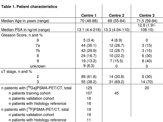
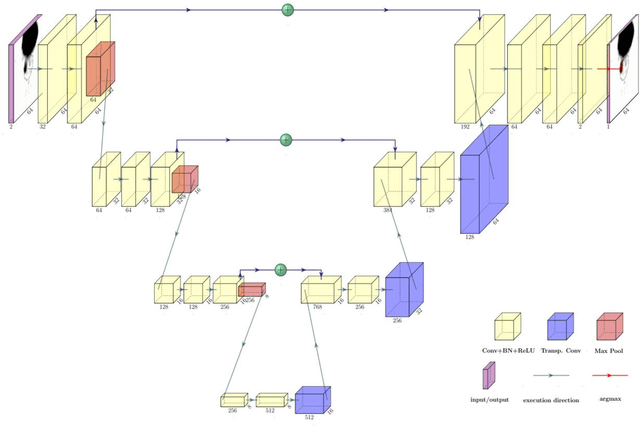
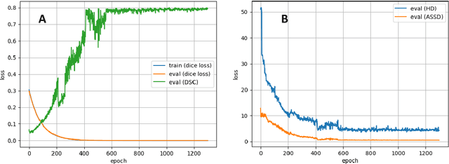
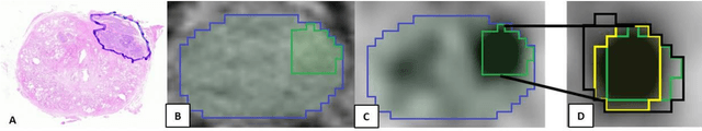
Accurate delineation of the intraprostatic gross tumour volume (GTV) is a prerequisite for treatment approaches in patients with primary prostate cancer (PCa). Prostate-specific membrane antigen positron emission tomography (PSMA-PET) may outperform MRI in GTV detection. However, visual GTV delineation underlies interobserver heterogeneity and is time consuming. The aim of this study was to develop a convolutional neural network (CNN) for automated segmentation of intraprostatic tumour (GTV-CNN) in PSMA-PET. Methods: The CNN (3D U-Net) was trained on [68Ga]PSMA-PET images of 152 patients from two different institutions and the training labels were generated manually using a validated technique. The CNN was tested on two independent internal (cohort 1: [68Ga]PSMA-PET, n=18 and cohort 2: [18F]PSMA-PET, n=19) and one external (cohort 3: [68Ga]PSMA-PET, n=20) test-datasets. Accordance between manual contours and GTV-CNN was assessed with Dice-S{\o}rensen coefficient (DSC). Sensitivity and specificity were calculated for the two internal test-datasets by using whole-mount histology. Results: Median DSCs for cohorts 1-3 were 0.84 (range: 0.32-0.95), 0.81 (range: 0.28-0.93) and 0.83 (range: 0.32-0.93), respectively. Sensitivities and specificities for GTV-CNN were comparable with manual expert contours: 0.98 and 0.76 (cohort 1) and 1 and 0.57 (cohort 2), respectively. Computation time was around 6 seconds for a standard dataset. Conclusion: The application of a CNN for automated contouring of intraprostatic GTV in [68Ga]PSMA- and [18F]PSMA-PET images resulted in a high concordance with expert contours and in high sensitivities and specificities in comparison with histology reference. This robust, accurate and fast technique may be implemented for treatment concepts in primary PCa. The trained model and the study's source code are available in an open source repository.