 Add to Chrome
 Add to Firefox
 Add to Edge
Cross-Modal Fusion of OCT and OCT angiography enface for Improved Diagnostics of Diabetic Retinopathy
Jul 04, 2026
Diabetic retinopathy (DR) is a leading cause of vision impairment worldwide, highlighting the need for accurate and accessible screening tools. Optical Coherence Tomography (OCT) provides high-resolution structural information of the retina, whereas OCT angiography (OCTA) offers complementary vascular information that is highly relevant for DR diagnosis. In this study, we propose a cross-modal fusion of OCT B-scans with single-channel en face OCTA using a bidirectional cross-modal attention network for automated DR classification. Two independent datasets, OCT500 and UIC, comprising 730 subjects in total, were utilized to evaluate performance under within-dataset, combined-dataset, and cross-dataset generalization settings. A ConvNeXt V2 model trained solely on OCT images served as the unimodal baseline. In addition to ground-truth (GT) OCTA, we explored the use of translated (TR) OCTA generated from OCT scans, eliminating the requirement for dedicated OCTA hardware. Experimental results demonstrate that cross-modal fusion consistently outperforms unimodal OCT classification across all evaluation scenarios. Fusion with GT OCTA improved classification accuracy and discriminative performance, while TR OCTA achieved comparable or superior results in most settings. Furthermore, TR OCTA improved sensitivity and cross-dataset generalization, indicating enhanced robustness to domain shifts. These findings demonstrate that attention-based OCT-OCTA en face fusion provides clinically meaningful improvements for DR detection and suggest that computationally generated OCTA can serve as a practical, low-cost alternative to hardware-acquired OCTA, enabling broader deployment of high-performance retinal screening systems in resource-limited clinical environments.

Quantitative Characterization of Retinal Features in Translated OCTA
Apr 24, 2024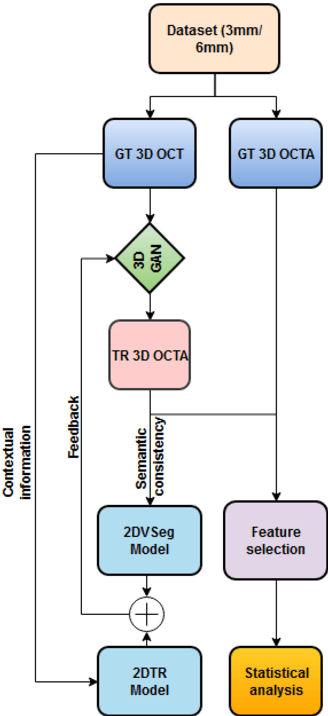
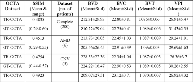
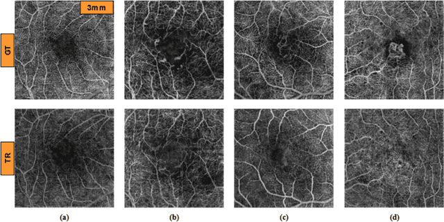
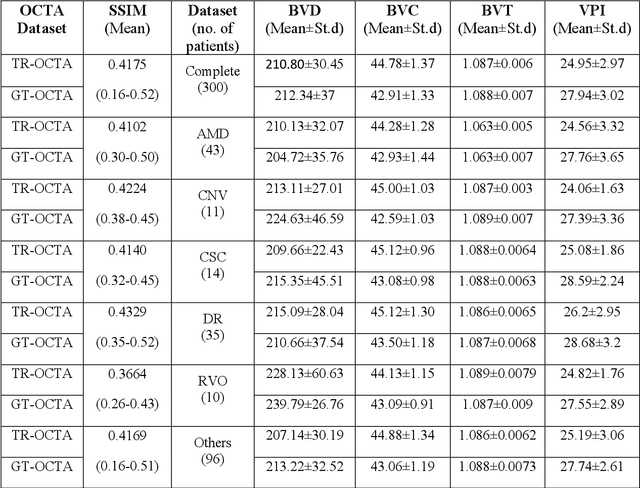
Purpose: This study explores the feasibility of using generative machine learning (ML) to translate Optical Coherence Tomography (OCT) images into Optical Coherence Tomography Angiography (OCTA) images, potentially bypassing the need for specialized OCTA hardware. Methods: The method involved implementing a generative adversarial network framework that includes a 2D vascular segmentation model and a 2D OCTA image translation model. The study utilizes a public dataset of 500 patients, divided into subsets based on resolution and disease status, to validate the quality of TR-OCTA images. The validation employs several quality and quantitative metrics to compare the translated images with ground truth OCTAs (GT-OCTA). We then quantitatively characterize vascular features generated in TR-OCTAs with GT-OCTAs to assess the feasibility of using TR-OCTA for objective disease diagnosis. Result: TR-OCTAs showed high image quality in both 3 and 6 mm datasets (high-resolution, moderate structural similarity and contrast quality compared to GT-OCTAs). There were slight discrepancies in vascular metrics, especially in diseased patients. Blood vessel features like tortuosity and vessel perimeter index showed a better trend compared to density features which are affected by local vascular distortions. Conclusion: This study presents a promising solution to the limitations of OCTA adoption in clinical practice by using vascular features from TR-OCTA for disease detection. Translation relevance: This study has the potential to significantly enhance the diagnostic process for retinal diseases by making detailed vascular imaging more widely available and reducing dependency on costly OCTA equipment.