 Add to Chrome
 Add to Firefox
 Add to Edge
VEELA: A Clinically-Constrained Benchmark for Liver Vessel Segmentation in Computed Tomography Angiography
May 21, 2026Accurate segmentation of hepatic and portal vessels in contrast-enhanced computed tomography angiography (CTA) remains challenging due to complex vascular topology, peripheral visibility limitations, and acquisition-induced ambiguities. While existing public datasets offer valuable benchmarks, few include clinically realistic annotation constraints. We introduce VEELA (Vessel Extraction and Extrication for Liver Analysis), a rigorously curated liver vessel dataset derived from 40 CTA scans inherited from the CHAOS grand-challenge cohort. All vessels were manually delineated slice-by-slice under multi-expert consensus, using a strict visibility-driven annotation policy and avoiding anatomically inferred interpolation. This design explicitly captures anatomical variability and imaging-related uncertainty. As a continuation of the CHAOS challenge, VEELA enables reproducible cross-benchmark evaluation while extending the scope to fine-grained hepatic and portal vessel segmentation. We further establish a standardized benchmarking framework and analyze complementary evaluation metrics, including topology-aware (clDice), overlap-based (IoU), boundary-sensitive (NSD), and geometry-aware (area, length) measures. Our results demonstrate that different metrics capture distinct aspects of vascular integrity, underscoring the necessity of multi-perspective evaluation for clinically meaningful vessel segmentation. VEELA is publicly released to facilitate reproducible research and support the development of robust vascular segmentation methods. Researchers can access the evaluation metrics, dataset, and submission platform at https://www.synapse.org/Synapse:syn65471967.
CHAOS Challenge -- Combined (CT-MR) Healthy Abdominal Organ Segmentation
Jan 17, 2020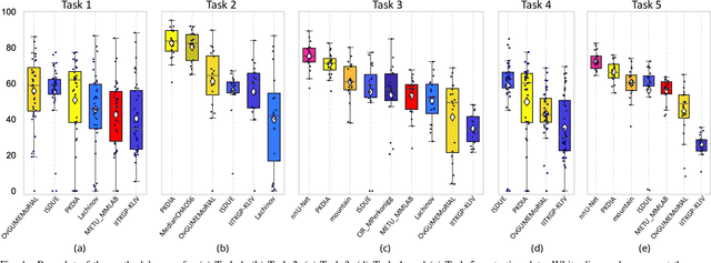
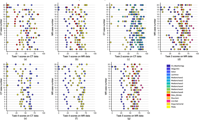
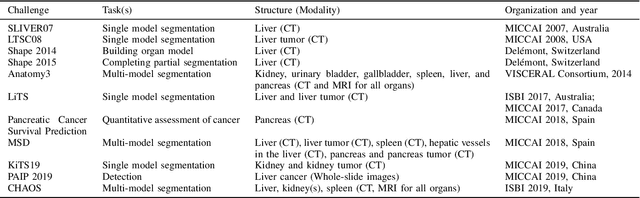
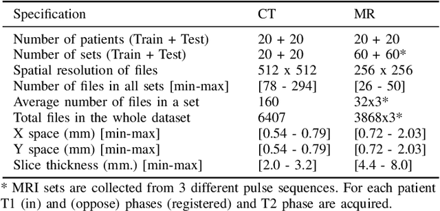
Segmentation of abdominal organs has been a comprehensive, yet unresolved, research field for many years. In the last decade, intensive developments in deep learning (DL) have introduced new state-of-the-art segmentation systems. Despite outperforming the overall accuracy of existing systems, the effects of DL model properties and parameters on the performance is hard to interpret. This makes comparative analysis a necessary tool to achieve explainable studies and systems. Moreover, the performance of DL for emerging learning approaches such as cross-modality and multi-modal tasks have been rarely discussed. In order to expand the knowledge in these topics, CHAOS -- Combined (CT-MR) Healthy Abdominal Organ Segmentation challenge has been organized in the IEEE International Symposium on Biomedical Imaging (ISBI), 2019, in Venice, Italy. Despite a large number of the previous abdomen related challenges, the majority of which are focused on tumor/lesion detection and/or classification with a single modality, CHAOS provides both abdominal CT and MR data from healthy subjects. Five different and complementary tasks have been designed to analyze the capabilities of the current approaches from multiple perspectives. The results are investigated thoroughly, compared with manual annotations and interactive methods. The outcomes are reported in detail to reflect the latest advancements in the field. CHAOS challenge and data will be available online to provide a continuous benchmark resource for segmentation.