 Add to Chrome
 Add to Firefox
 Add to Edge
Automatic brain tissue segmentation in fetal MRI using convolutional neural networks
Jun 11, 2019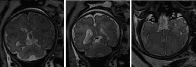
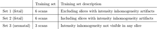
MR images of fetuses allow clinicians to detect brain abnormalities in an early stage of development. The cornerstone of volumetric and morphologic analysis in fetal MRI is segmentation of the fetal brain into different tissue classes. Manual segmentation is cumbersome and time consuming, hence automatic segmentation could substantially simplify the procedure. However, automatic brain tissue segmentation in these scans is challenging owing to artifacts including intensity inhomogeneity, caused in particular by spontaneous fetal movements during the scan. Unlike methods that estimate the bias field to remove intensity inhomogeneity as a preprocessing step to segmentation, we propose to perform segmentation using a convolutional neural network that exploits images with synthetically introduced intensity inhomogeneity as data augmentation. The method first uses a CNN to extract the intracranial volume. Thereafter, another CNN with the same architecture is employed to segment the extracted volume into seven brain tissue classes: cerebellum, basal ganglia and thalami, ventricular cerebrospinal fluid, white matter, brain stem, cortical gray matter and extracerebral cerebrospinal fluid. To make the method applicable to slices showing intensity inhomogeneity artifacts, the training data was augmented by applying a combination of linear gradients with random offsets and orientations to image slices without artifacts.
Generative adversarial network for segmentation of motion affected neonatal brain MRI
Jun 11, 2019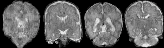
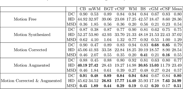
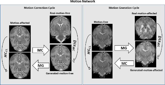
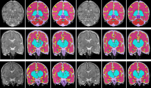
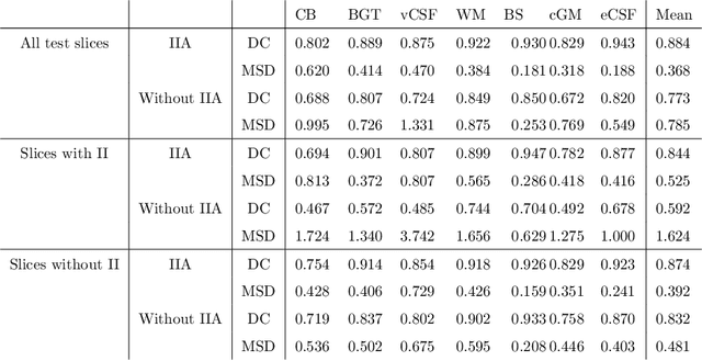
Automatic neonatal brain tissue segmentation in preterm born infants is a prerequisite for evaluation of brain development. However, automatic segmentation is often hampered by motion artifacts caused by infant head movements during image acquisition. Methods have been developed to remove or minimize these artifacts during image reconstruction using frequency domain data. However, frequency domain data might not always be available. Hence, in this study we propose a method for removing motion artifacts from the already reconstructed MR scans. The method employs a generative adversarial network trained with a cycle consistency loss to transform slices affected by motion into slices without motion artifacts, and vice versa. In the experiments 40 T2-weighted coronal MR scans of preterm born infants imaged at 30 weeks postmenstrual age were used. All images contained slices affected by motion artifacts hampering automatic tissue segmentation. To evaluate whether correction allows more accurate image segmentation, the images were segmented into 8 tissue classes: cerebellum, myelinated white matter, basal ganglia and thalami, ventricular cerebrospinal fluid, white matter, brain stem, cortical gray matter, and extracerebral cerebrospinal fluid. Images corrected for motion and corresponding segmentations were qualitatively evaluated using 5-point Likert scale. Before the correction of motion artifacts, median image quality and quality of corresponding automatic segmentations were assigned grade 2 (poor) and 3 (moderate), respectively. After correction of motion artifacts, both improved to grades 3 and 4, respectively. The results indicate that correction of motion artifacts in the image space using the proposed approach allows accurate segmentation of brain tissue classes in slices affected by motion artifacts.
Automatic segmentation of the intracranialvolume in fetal MR images
Jul 31, 2017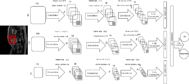
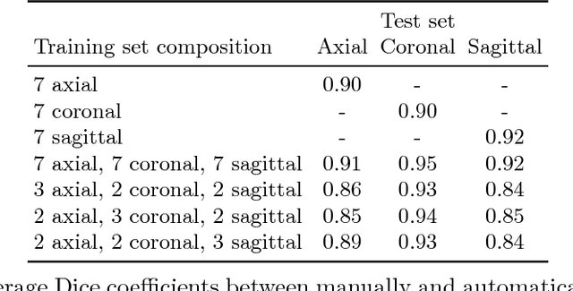
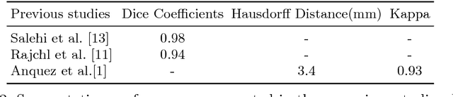
MR images of the fetus allow non-invasive analysis of the fetal brain. Quantitative analysis of fetal brain development requires automatic brain tissue segmentation that is typically preceded by segmentation of the intracranial volume (ICV). This is challenging because fetal MR images visualize the whole moving fetus and in addition partially visualize the maternal body. This paper presents an automatic method for segmentation of the ICV in fetal MR images. The method employs a multi-scale convolutional neural network in 2D slices to enable learning spatial information from larger context as well as detailed local information. The method is developed and evaluated with 30 fetal T2-weighted MRI scans (average age $33.2\pm1.2$ weeks postmenstrual age). The set contains $10$ scans acquired in axial, $10$ in coronal and $10$ in sagittal imaging planes. A reference standard was defined in all images by manual annotation of the intracranial volume in $10$ equidistantly distributed slices. The automatic analysis was performed by training and testing the network using scans acquired in the representative imaging plane as well as combining the training data from all imaging planes. On average, the automatic method achieved Dice coefficients of 0.90 for the axial images, 0.90 for the coronal images and 0.92 for the sagittal images. Combining the training sets resulted in average Dice coefficients of 0.91 for the axial images, 0.95 for the coronal images, and 0.92 for the sagittal images. The results demonstrate that the evaluated method achieved good performance in extracting ICV in fetal MR scans regardless of the imaging plane.