 Add to Chrome
 Add to Firefox
 Add to Edge
Multiple Instance Learning with random sampling for Whole Slide Image Classification
Mar 08, 2024In computational pathology, random sampling of patches during training of Multiple Instance Learning (MIL) methods is computationally efficient and serves as a regularization strategy. Despite its promising benefits, questions concerning performance trends for varying sample sizes and its influence on model interpretability remain. Addressing these, we reach an optimal performance enhancement of 1.7% using thirty percent of patches on the CAMELYON16 dataset, and 3.7% with only eight samples on the TUPAC16 dataset. We also find interpretability effects are strongly dataset-dependent, with interpretability impacted on CAMELYON16, while remaining unaffected on TUPAC16. This reinforces that both the performance and interpretability relationships with sampling are closely task-specific. End-to-end training with 1024 samples reveals improvements across both datasets compared to pre-extracted features, further highlighting the potential of this efficient approach.
Automatic segmentation of the intracranialvolume in fetal MR images
Jul 31, 2017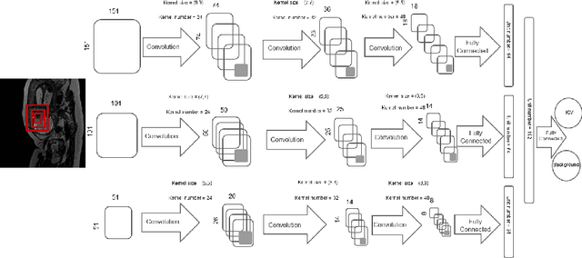
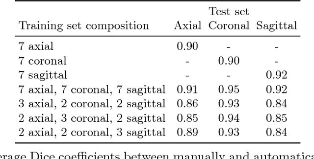
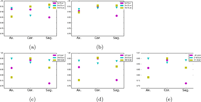
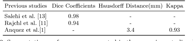
MR images of the fetus allow non-invasive analysis of the fetal brain. Quantitative analysis of fetal brain development requires automatic brain tissue segmentation that is typically preceded by segmentation of the intracranial volume (ICV). This is challenging because fetal MR images visualize the whole moving fetus and in addition partially visualize the maternal body. This paper presents an automatic method for segmentation of the ICV in fetal MR images. The method employs a multi-scale convolutional neural network in 2D slices to enable learning spatial information from larger context as well as detailed local information. The method is developed and evaluated with 30 fetal T2-weighted MRI scans (average age $33.2\pm1.2$ weeks postmenstrual age). The set contains $10$ scans acquired in axial, $10$ in coronal and $10$ in sagittal imaging planes. A reference standard was defined in all images by manual annotation of the intracranial volume in $10$ equidistantly distributed slices. The automatic analysis was performed by training and testing the network using scans acquired in the representative imaging plane as well as combining the training data from all imaging planes. On average, the automatic method achieved Dice coefficients of 0.90 for the axial images, 0.90 for the coronal images and 0.92 for the sagittal images. Combining the training sets resulted in average Dice coefficients of 0.91 for the axial images, 0.95 for the coronal images, and 0.92 for the sagittal images. The results demonstrate that the evaluated method achieved good performance in extracting ICV in fetal MR scans regardless of the imaging plane.