 Add to Chrome
 Add to Firefox
 Add to Edge
PR3DICTR: A modular AI framework for medical 3D image-based detection and outcome prediction
Apr 03, 2026Three-dimensional medical image data and computer-aided decision making, particularly using deep learning, are becoming increasingly important in the medical field. To aid in these developments we introduce PR3DICTR: Platform for Research in 3D Image Classification and sTandardised tRaining. Built using community-standard distributions (PyTorch and MONAI), PR3DICTR provides an open-access, flexible and convenient framework for prediction model development, with an explicit focus on classification using three-dimensional medical image data. By combining modular design principles and standardization, it aims to alleviate developmental burden whilst retaining adjustability. It provides users with a wealth of pre-established functionality, for instance in model architecture design options, hyper-parameter solutions and training methodologies, but still gives users the opportunity and freedom to ``plug in'' their own solutions or modules. PR3DICTR can be applied to any binary or event-based three-dimensional classification task and can work with as little as two lines of code.
Corneal Pachymetry by AS-OCT after Descemet's Membrane Endothelial Keratoplasty
Feb 15, 2021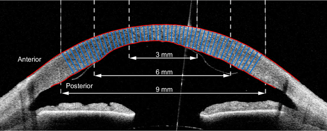
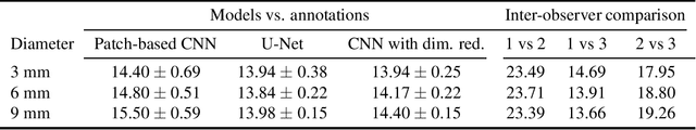
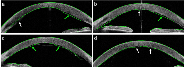
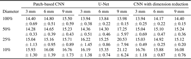
Corneal thickness (pachymetry) maps can be used to monitor restoration of corneal endothelial function, for example after Descemet's membrane endothelial keratoplasty (DMEK). Automated delineation of the corneal interfaces in anterior segment optical coherence tomography (AS-OCT) can be challenging for corneas that are irregularly shaped due to pathology, or as a consequence of surgery, leading to incorrect thickness measurements. In this research, deep learning is used to automatically delineate the corneal interfaces and measure corneal thickness with high accuracy in post-DMEK AS-OCT B-scans. Three different deep learning strategies were developed based on 960 B-scans from 68 patients. On an independent test set of 320 B-scans, corneal thickness could be measured with an error of 13.98 to 15.50 micrometer for the central 9 mm range, which is less than 3% of the average corneal thickness. The accurate thickness measurements were used to construct detailed pachymetry maps. Moreover, follow-up scans could be registered based on anatomical landmarks to obtain differential pachymetry maps. These maps may enable a more comprehensive understanding of the restoration of the endothelial function after DMEK, where thickness often varies throughout different regions of the cornea, and subsequently contribute to a standardized postoperative regime.