 Add to Chrome
 Add to Firefox
 Add to Edge
SPARC: Slice-to-volume Pipeline for Automated Reconstruction of gated 3D+time fetal Cardiac MRI
Aug 19, 2026
Fetal cardiac MRI (fCMR) provides valuable diagnostic information complementary to echocardiography, particularly for complex congenital heart disease (CHD). Dynamic cine imaging captures cardiac motion essential for assessment of cardiac function; however, the reconstruction of 3D+time cine volumes from 2D+time acquired slices remains challenging due to unpredictable fetal motion and the absence of automated and robust processing tools suitable for clinical deployment. We present the SPARC pipeline (Slice-to-volume Pipeline for Automated Reconstruction of gated 3D+time fetal Cardiac MRI) which combines physics-informed slice-to-volume reconstruction (SVR) of Doppler ultrasound (DUS) gated stacks of slices, assisted by deep learning (DL) models for thoracic segmentation and anatomical reorientation. The proposed SVR algorithm achieves a tenfold reduction in reconstruction time relative to existing frame-wise approaches ($4.8 \pm 1.0$ vs $49.0 \pm 14.1$ min, $p < 0.0001$) while improving the reconstruction quality. Thoracic segmentation performance using ensemble aggregation exceeded inter-rater agreement (Dice $84.7 \pm 3.9\%$ vs $81.4 \pm 7.7\%$, $p<0.05$), while anatomical reorientation achieved a success rate of $90.1\%$. End-to-end evaluation on a large held-out clinical cohort ($n = 121$) demonstrated fully automatic processing in $82.6\%$ of cases with a mean runtime of $7.1 \pm 1.3$ min, compatible with clinical deployment. The complete SPARC pipeline is publicly available as a Docker container https://hub.docker.com/r/aboutill/sparc and is currently deployed at our institution as a clinical research tool.

Multi-task learning for joint weakly-supervised segmentation and aortic arch anomaly classification in fetal cardiac MRI
Nov 13, 2023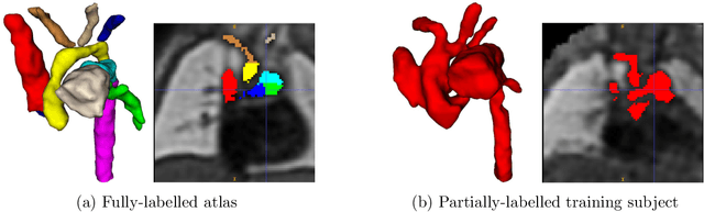

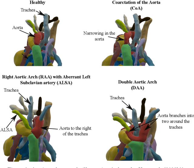
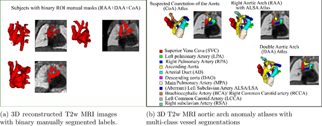
Congenital Heart Disease (CHD) is a group of cardiac malformations present already during fetal life, representing the prevailing category of birth defects globally. Our aim in this study is to aid 3D fetal vessel topology visualisation in aortic arch anomalies, a group which encompasses a range of conditions with significant anatomical heterogeneity. We present a multi-task framework for automated multi-class fetal vessel segmentation from 3D black blood T2w MRI and anomaly classification. Our training data consists of binary manual segmentation masks of the cardiac vessels' region in individual subjects and fully-labelled anomaly-specific population atlases. Our framework combines deep learning label propagation using VoxelMorph with 3D Attention U-Net segmentation and DenseNet121 anomaly classification. We target 11 cardiac vessels and three distinct aortic arch anomalies, including double aortic arch, right aortic arch, and suspected coarctation of the aorta. We incorporate an anomaly classifier into our segmentation pipeline, delivering a multi-task framework with the primary motivation of correcting topological inaccuracies of the segmentation. The hypothesis is that the multi-task approach will encourage the segmenter network to learn anomaly-specific features. As a secondary motivation, an automated diagnosis tool may have the potential to enhance diagnostic confidence in a decision support setting. Our results showcase that our proposed training strategy significantly outperforms label propagation and a network trained exclusively on propagated labels. Our classifier outperforms a classifier trained exclusively on T2w volume images, with an average balanced accuracy of 0.99 (0.01) after joint training. Adding a classifier improves the anatomical and topological accuracy of all correctly classified double aortic arch subjects.
* Accepted for publication at the Journal of Machine Learning for Biomedical Imaging (MELBA) https://melba-journal.org/2023:015