 Add to Chrome
 Add to Firefox
 Add to Edge
SPARC: Slice-to-volume Pipeline for Automated Reconstruction of gated 3D+time fetal Cardiac MRI
Aug 19, 2026
Fetal cardiac MRI (fCMR) provides valuable diagnostic information complementary to echocardiography, particularly for complex congenital heart disease (CHD). Dynamic cine imaging captures cardiac motion essential for assessment of cardiac function; however, the reconstruction of 3D+time cine volumes from 2D+time acquired slices remains challenging due to unpredictable fetal motion and the absence of automated and robust processing tools suitable for clinical deployment. We present the SPARC pipeline (Slice-to-volume Pipeline for Automated Reconstruction of gated 3D+time fetal Cardiac MRI) which combines physics-informed slice-to-volume reconstruction (SVR) of Doppler ultrasound (DUS) gated stacks of slices, assisted by deep learning (DL) models for thoracic segmentation and anatomical reorientation. The proposed SVR algorithm achieves a tenfold reduction in reconstruction time relative to existing frame-wise approaches ($4.8 \pm 1.0$ vs $49.0 \pm 14.1$ min, $p < 0.0001$) while improving the reconstruction quality. Thoracic segmentation performance using ensemble aggregation exceeded inter-rater agreement (Dice $84.7 \pm 3.9\%$ vs $81.4 \pm 7.7\%$, $p<0.05$), while anatomical reorientation achieved a success rate of $90.1\%$. End-to-end evaluation on a large held-out clinical cohort ($n = 121$) demonstrated fully automatic processing in $82.6\%$ of cases with a mean runtime of $7.1 \pm 1.3$ min, compatible with clinical deployment. The complete SPARC pipeline is publicly available as a Docker container https://hub.docker.com/r/aboutill/sparc and is currently deployed at our institution as a clinical research tool.

Unsupervised self-organising map of prostate cell Raman spectra shows disease-state subclustering
Mar 12, 2024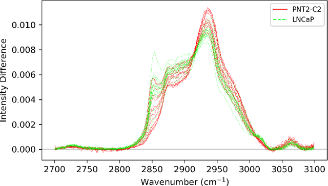
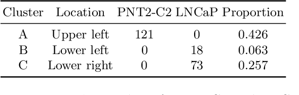
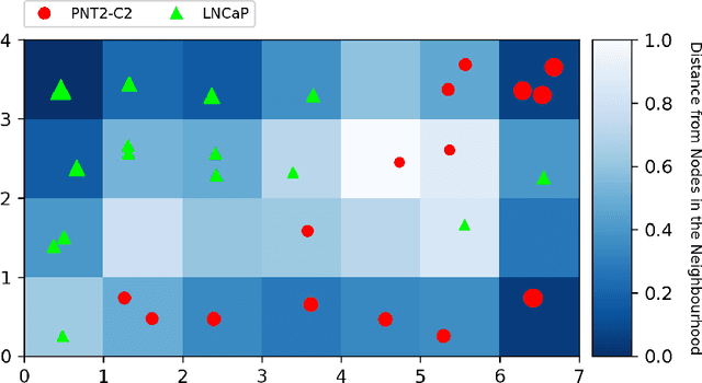
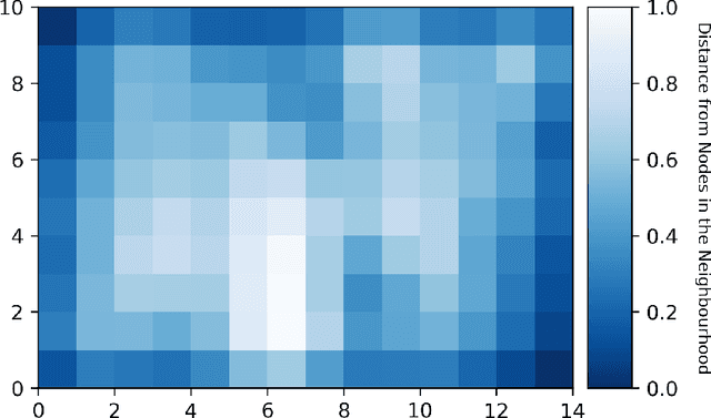
Prostate cancer is a disease which poses an interesting clinical question: should it be treated? A small subset of prostate cancers are aggressive and require removal and treatment to prevent metastatic spread. However, conventional diagnostics remain challenged to risk-stratify such patients, hence, new methods of approach to biomolecularly subclassify the disease are needed. Here we use an unsupervised, self-organising map approach to analyse live-cell Raman spectroscopy data obtained from prostate cell-lines; our aim is to test the feasibility of this method to differentiate, at the single-cell-level, cancer from normal using high-dimensional datasets with minimal preprocessing. The results demonstrate not only successful separation of normal prostate and cancer cells, but also a new subclustering of the prostate cancer cell-line into two groups. Initial analysis of the spectra from each of the cancer subclusters demonstrates a differential expression of lipids, which, against the normal control, may be linked to disease-related changes in cellular signalling.