 Add to Chrome
 Add to Firefox
 Add to Edge
Variable Resolution Sampling and Deep Learning Image Recovery for Accelerated Multi-Spectral MRI Near Metal Implants
Oct 30, 2024Purpose: This study presents a variable resolution (VR) sampling and deep learning reconstruction approach for multi-spectral MRI near metal implants, aiming to reduce scan times while maintaining image quality. Background: The rising use of metal implants has increased MRI scans affected by metal artifacts. Multi-spectral imaging (MSI) reduces these artifacts but sacrifices acquisition efficiency. Methods: This retrospective study on 1.5T MSI knee and hip data from patients with metal hardware used a novel spectral undersampling scheme to improve acquisition efficiency by ~40%. U-Net-based deep learning models were trained for reconstruction. Image quality was evaluated using SSIM, PSNR, and RESI metrics. Results: Deep learning reconstructions of undersampled VR data (DL-VR) showed significantly higher SSIM and PSNR values (p<0.001) compared to conventional reconstruction (CR-VR), with improved edge sharpness. Edge sharpness in DL-reconstructed images matched fully sampled references (p=0.5). Conclusion: This approach can potentially enhance MRI examinations near metal implants by reducing scan times or enabling higher resolution. Further prospective studies are needed to assess clinical value.
On Functional Activations in Deep Neural Networks
Nov 17, 2023


Background: Deep neural networks have proven to be powerful computational tools for modeling, prediction, and generation. However, the workings of these models have generally been opaque. Recent work has shown that the performance of some models are modulated by overlapping functional networks of connections within the models. Here the techniques of functional neuroimaging are applied to an exemplary large language model to probe its functional structure. Methods: A series of block-designed task-based prompt sequences were generated to probe the Facebook Galactica-125M model. Tasks included prompts relating to political science, medical imaging, paleontology, archeology, pathology, and random strings presented in an off/on/off pattern with prompts about other random topics. For the generation of each output token, all layer output values were saved to create an effective time series. General linear models were fit to the data to identify layer output values which were active with the tasks. Results: Distinct, overlapping networks were identified with each task. Most overlap was observed between medical imaging and pathology networks. These networks were repeatable across repeated performance of related tasks, and correspondence of identified functional networks and activation in tasks not used to define the functional networks was shown to accurately identify the presented task. Conclusion: The techniques of functional neuroimaging can be applied to deep neural networks as a means to probe their workings. Identified functional networks hold the potential for use in model alignment, modulation of model output, and identifying weights to target in fine-tuning.
Deep-learning based Tools for Automated Protocol Definition of Advanced Diagnostic Imaging Exams
May 28, 2021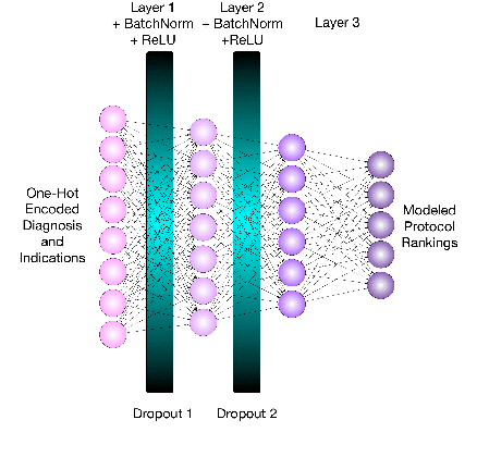
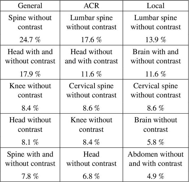
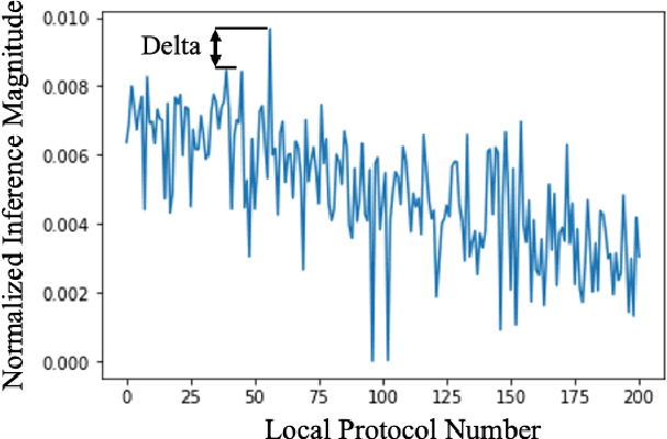
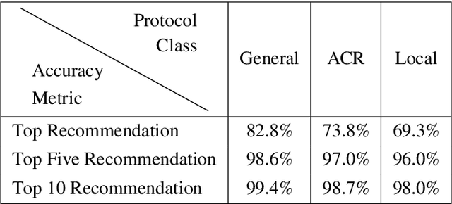
Purpose: This study evaluates the effectiveness and impact of automated order-based protocol assignment for magnetic resonance imaging (MRI) exams using natural language processing (NLP) and deep learning (DL). Methods: NLP tools were applied to retrospectively process orders from over 116,000 MRI exams with 200 unique sub-specialized protocols ("Local" protocol class). Separate DL models were trained on 70\% of the processed data for "Local" protocols as well as 93 American College of Radiology ("ACR") protocols and 48 "General" protocols. The DL Models were assessed in an "auto-protocoling (AP)" inference mode which returns the top recommendation and in a "clinical decision support (CDS)" inference mode which returns up to 10 protocols for radiologist review. The accuracy of each protocol recommendation was computed and analyzed based on the difference between the normalized output score of the corresponding neural net for the top two recommendations. Results: The top predicted protocol in AP mode was correct for 82.8%, 73.8%, and 69.3% of the test cases for "General", "ACR", and "Local" protocol classes, respectively. Higher levels of accuracy over 96% were obtained for all protocol classes in CDS mode. However, at current validation performance levels, the proposed models offer modest, positive, financial impact on large-scale imaging networks. Conclusions: DL-based protocol automation is feasible and can be tuned to route substantial fractions of exams for auto-protocoling, with higher accuracy with more general protocols. Economic analyses of the tested algorithms indicate that improved algorithm performance is required to yield a practical exam auto-protocoling tool for sub-specialized imaging exams.
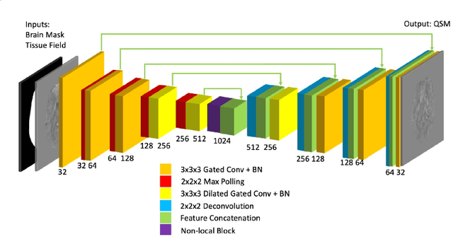
MRI Tissue Magnetism Quantification through Total Field Inversion with Deep Neural Networks
Apr 11, 2019


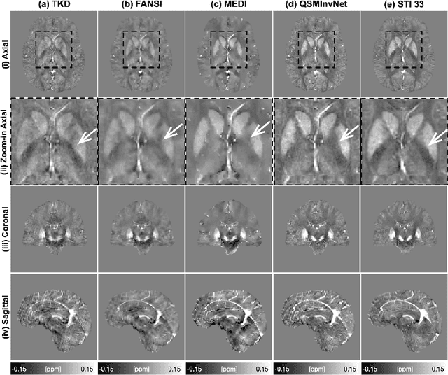
Quantitative susceptibility mapping (QSM) utilizes MRI signal phase to infer estimates of local tissue magnetism (magnetic susceptibility), which has been shown useful to provide novel image contrast and as biomarkers of abnormal tissue. QSM requires addressing a challenging post-processing problem: filtering of image phase estimates and inversion of the phase to susceptibility relationship. A wide variety of quantification errors, robustness limitations, and artifacts plague QSM algorithms. To overcome these limitations, a robust deep-learning-based single-step QSM reconstruction approach is proposed and demonstrated. This neural network was trained using magnetostatic physics simulations based on in-vivo data sources. Random perturbations were added to the physics simulations to provide sufficient input-label pairs for the training purposes. The network was quantitatively tested using gold-standard in-silico labeled datasets against established QSM total field inversion approaches. In addition, the algorithm was applied to susceptibility-weighted imaging (SWI) data collected on a cohort of clinical subjects with brain hemmhorage. When quantitatively compared against gold-standard in-silico labels, the proposed algorithm outperformed the existing comparable approaches. High quality QSM were consistently estimated from clinical susceptibility-weighted data on 100 subjects without any noticeable inversion failures. The proposed approach was able to robustly generate high quality QSM with improved accuracy in in-silico gold-standard experiments. QSM produced by the proposed method can be generated in real-time on existing MRI scanner platforms and provide enhanced visualization and quantification of magnetism-based tissue contrasts.
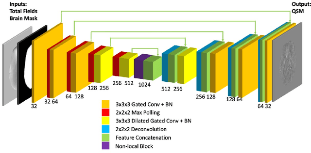
Non-locally Encoder-Decoder Convolutional Network for Whole Brain QSM Inversion
Apr 11, 2019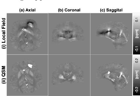
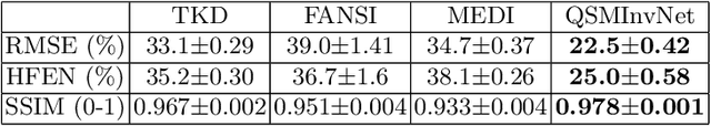
Quantitative Susceptibility Mapping (QSM) reconstruction is a challenging inverse problem driven by ill conditioning of its field-to -susceptibility transformation. State-of-art QSM reconstruction methods either suffer from image artifacts or long computation times, which limits QSM clinical translation efforts. To overcome these limitations, a non-locally encoder-decoder gated convolutional neural network is trained to infer whole brain susceptibility map, using the local field and brain mask as the inputs. The performance of the proposed method is evaluated relative to synthetic data, a publicly available challenge dataset, and clinical datasets. The proposed approach can outperform existing methods on quantitative metrics and visual assessment of image sharpness and streaking artifacts. The estimated susceptibility maps can preserve conspicuity of fine features and suppress streaking artifacts. The demonstrated methods have potential value in advancing QSM clinical research and aiding in the translation of QSM to clinical operations.