 Add to Chrome
 Add to Firefox
 Add to Edge
Reversible Image Watermarking for Health Informatics Systems Using Distortion Compensation in Wavelet Domain
Feb 21, 2018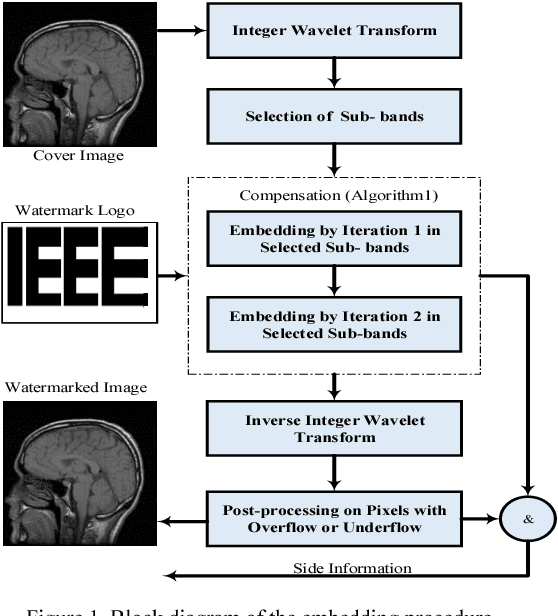
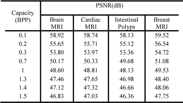
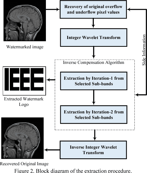
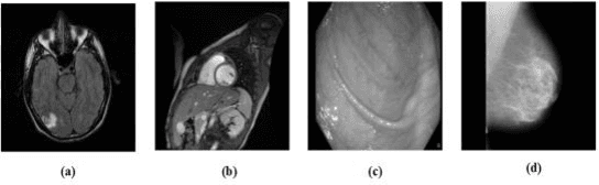
Reversible image watermarking guaranties restoration of both original cover and watermark logo from the watermarked image. Capacity and distortion of the image under reversible watermarking are two important parameters. In this study a reversible watermarking is investigated with focusing on increasing the embedding capacity and reducing the distortion in medical images. Integer wavelet transform is used for embedding where in each iteration, one watermark bit is embedded in one transform coefficient. We devise a novel approach that when a coefficient is modified in an iteration, the produced distortion is compensated in the next iteration. This distortion compensation method would result in low distortion rate. The proposed method is tested on four types of medical images including MRI of brain, cardiac MRI, MRI of breast, and intestinal polyp images. Using a one-level wavelet transform, maximum capacity of 1.5 BPP is obtained. Experimental results demonstrate that the proposed method is superior to the state-of-the-art works in terms of capacity and distortion.
Lossless Image Compression Algorithm for Wireless Capsule Endoscopy by Content-Based Classification of Image Blocks
Feb 21, 2018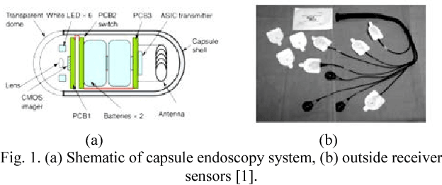
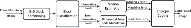
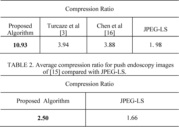
Recent advances in capsule endoscopy systems have introduced new methods and capabilities. The capsule endoscopy system, by observing the entire digestive tract, has significantly improved diagnosing gastrointestinal disorders and diseases. The system has challenges such as the need to enhance the quality of the transmitted images, low frame rates of transmission, and battery lifetime that need to be addressed. One of the important parts of a capsule endoscopy system is the image compression unit. Better compression of images increases the frame rate and hence improves the diagnosis process. In this paper a high precision compression algorithm with high compression ratio is proposed. In this algorithm we use the similarity between frames to compress the data more efficiently.
Left Ventricle Segmentation in Cardiac MR Images Using Fully Convolutional Network
Feb 21, 2018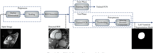
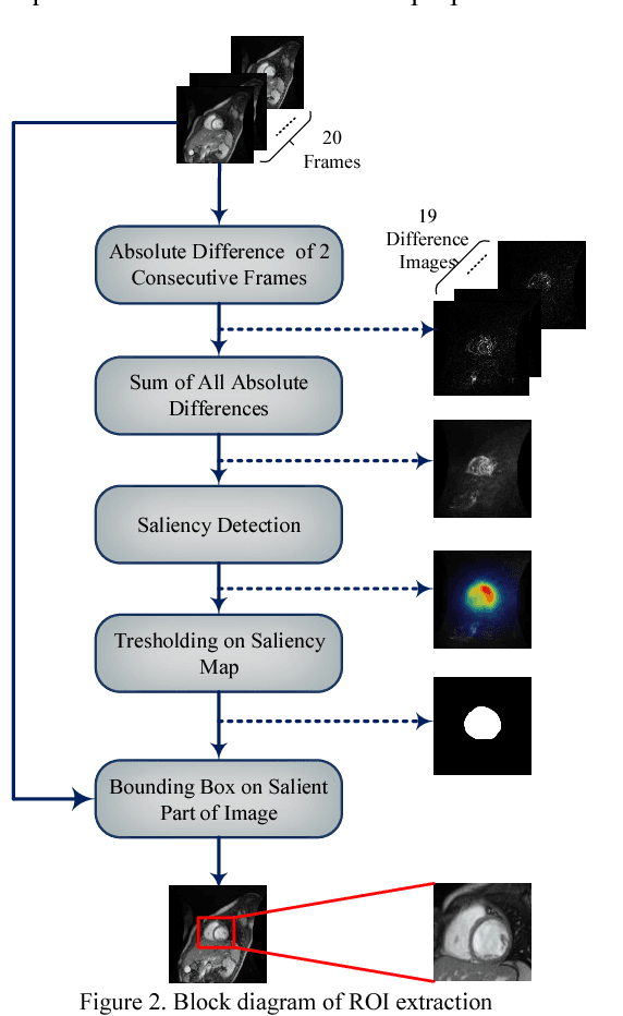
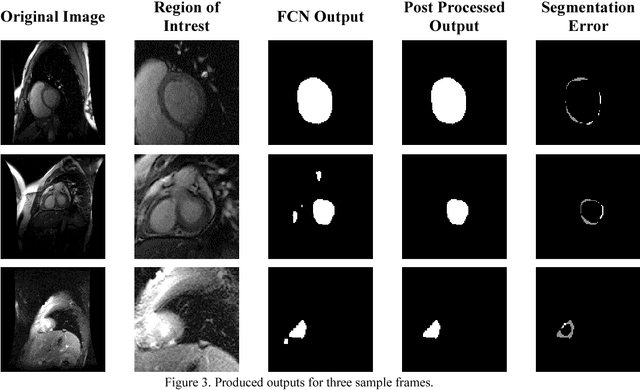
Medical image analysis, especially segmenting a specific organ, has an important role in developing clinical decision support systems. In cardiac magnetic resonance (MR) imaging, segmenting the left and right ventricles helps physicians diagnose different heart abnormalities. There are challenges for this task, including the intensity and shape similarity between left ventricle and other organs, inaccurate boundaries and presence of noise in most of the images. In this paper we propose an automated method for segmenting the left ventricle in cardiac MR images. We first automatically extract the region of interest, and then employ it as an input of a fully convolutional network. We train the network accurately despite the small number of left ventricle pixels in comparison with the whole image. Thresholding on the output map of the fully convolutional network and selection of regions based on their roundness are performed in our proposed post-processing phase. The Dice score of our method reaches 87.24% by applying this algorithm on the York dataset of heart images.
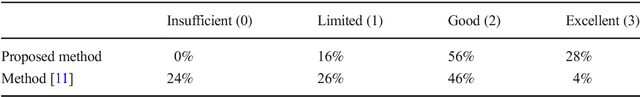
Lossless Compression of Angiogram Foreground with Visual Quality Preservation of Background
Feb 21, 2018
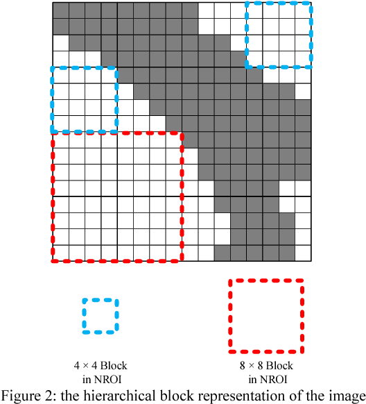
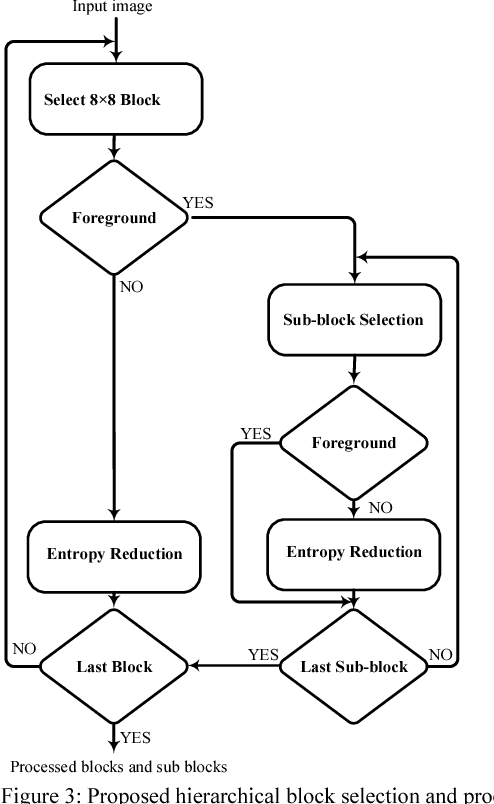
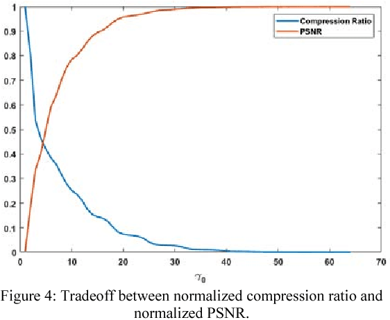
By increasing the volume of telemedicine information, the need for medical image compression has become more important. In angiographic images, a small ratio of the entire image usually belongs to the vasculature that provides crucial information for diagnosis. Other parts of the image are diagnostically less important and can be compressed with higher compression ratio. However, the quality of those parts affect the visual perception of the image as well. Existing methods compress foreground and background of angiographic images using different techniques. In this paper we first utilize convolutional neural network to segment vessels and then represent a hierarchical block processing algorithm capable of both eliminating the background redundancies and preserving the overall visual quality of angiograms.
Adaptive Real-Time Removal of Impulse Noise in Medical Images
Oct 12, 2017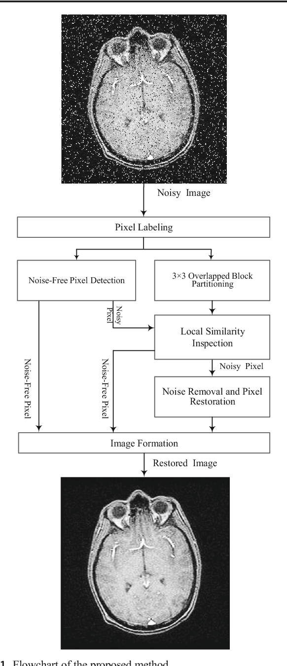
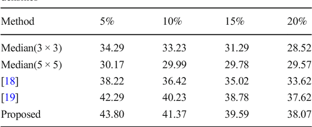
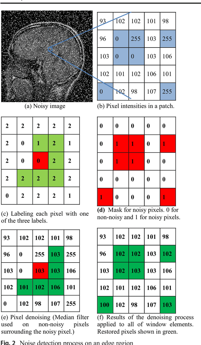
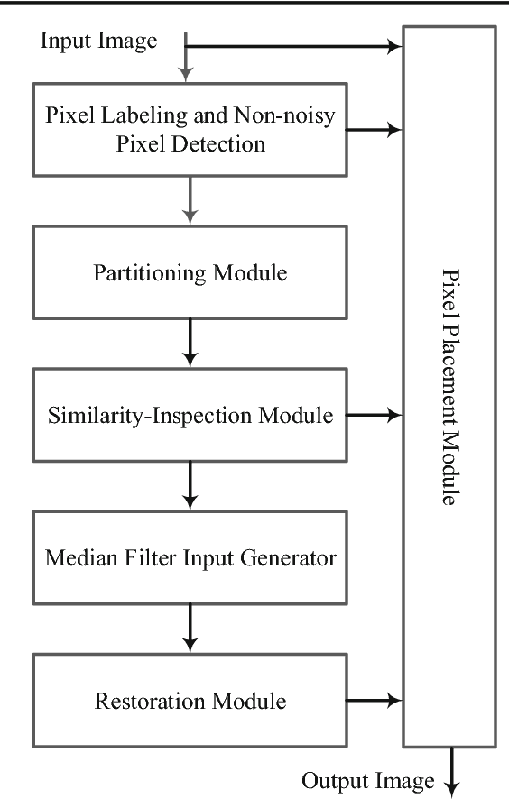
Noise is an important factor that degrades the quality of medical images. Impulse noise is a common noise, which is caused by malfunctioning of sensor elements or errors in the transmission of images. In medical images due to presence of white foreground and black background, many pixels have intensities similar to impulse noise and distinction between noisy and regular pixels is difficult. In software techniques, the accuracy of the noise removal is more important than the algorithm's complexity. But for hardware implementation having a low complexity algorithm with an acceptable accuracy is essential. In this paper a low complexity de-noising method is proposed that removes the noise by local analysis of the image blocks. The proposed method distinguishes non-noisy pixels that have noise-like intensities. All steps are designed to have low hardware complexity. Simulation results show that for different magnetic resonance images, the proposed method removes impulse noise with an acceptable accuracy.
Vessel Segmentation and Catheter Detection in X-Ray Angiograms Using Superpixels
Sep 08, 2017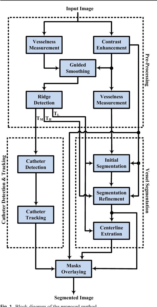
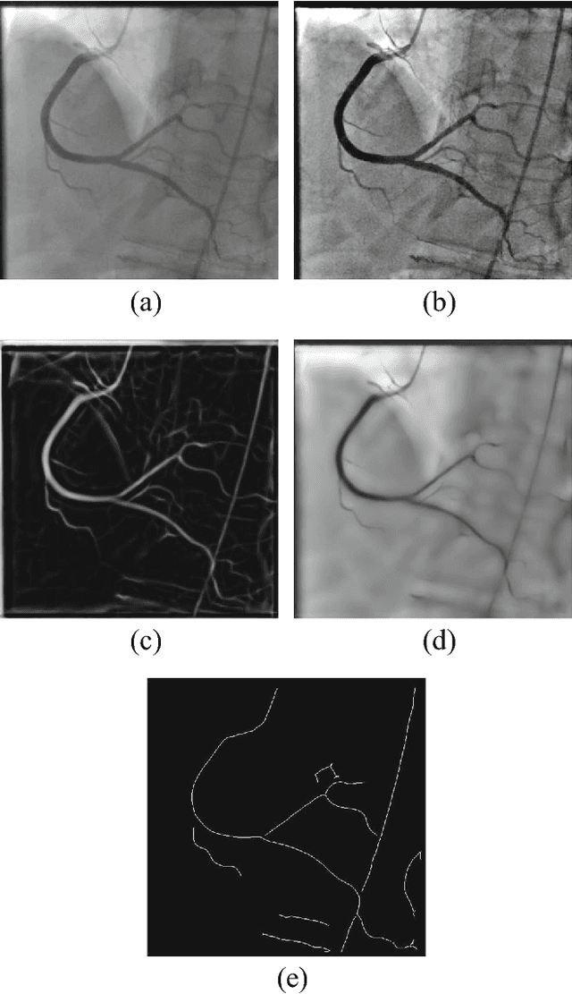

Coronary artery disease (CAD) is the leading causes of death around the world. One of the most common imaging methods for diagnosing this disease is X-ray angiography. Diagnosing using these images is usually challenging due to non-uniform illumination, low contrast, presence of other body tissues, presence of catheter etc. These challenges make the diagnoses task of cardiologists tougher and more prone to misdiagnosis. In this paper we propose a new automated framework for coronary arteries segmentation, catheter detection and center-line extraction in x-ray angiography images. Our proposed segmentation method is based on superpixels. In this method at first three different superpixel scales are exploited and a measure for vesselness probability of each superpixel is determined. A majority voting is used for obtaining an initial segmentation map from these three superpixel scales. This initial segmentation is refined by finding the orthogonal line on each ridge pixel of vessel region. In this framework we use our catheter detection and tracking method which detects the catheter by finding its ridge in the first frame and traces in other frames by fitting a second order polynomial on it. Also we use the image ridges for extracting the coronary arteries centerlines. We evaluated our method qualitatively and quantitatively on two different challenging datasets and compared it with one of the previous well-known coronary arteries segmentation methods. Our method could detect the catheter and reduced the false positive rate in addition to achieving better segmentation results. The evaluation results prove that our method performs better in a much shorter time.
Hand Gesture Recognition for Contactless Device Control in Operating Rooms
Nov 13, 2016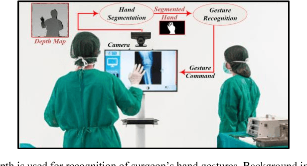
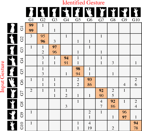
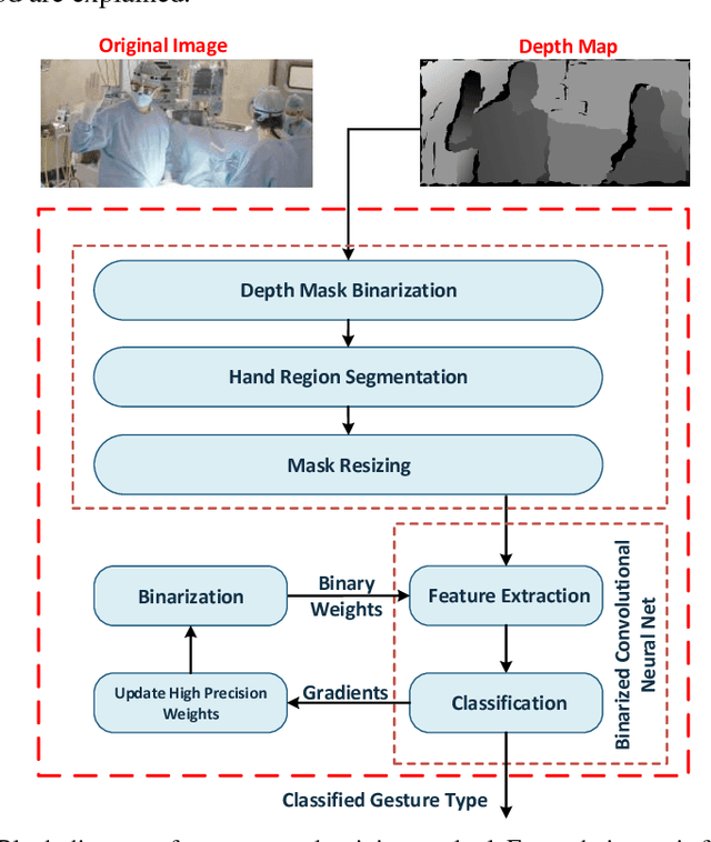

Hand gesture is one of the most important means of touchless communication between human and machines. There is a great interest for commanding electronic equipment in surgery rooms by hand gesture for reducing the time of surgery and the potential for infection. There are challenges in implementation of a hand gesture recognition system. It has to fulfill requirements such as high accuracy and fast response. In this paper we introduce a system of hand gesture recognition based on a deep learning approach. Deep learning is known as an accurate detection model, but its high complexity prevents it from being fabricated as an embedded system. To cope with this problem, we applied some changes in the structure of our work to achieve low complexity. As a result, the proposed method could be implemented on a naive embedded system. Our experiments show that the proposed system results in higher accuracy while having less complexity in comparison with the existing comparable methods.
Extraction of Skin Lesions from Non-Dermoscopic Images Using Deep Learning
Sep 08, 2016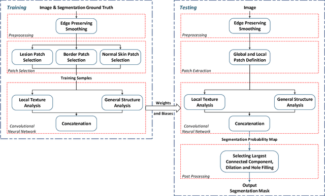
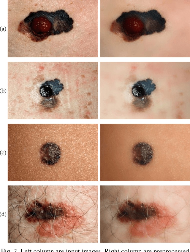
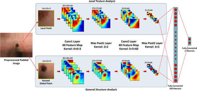
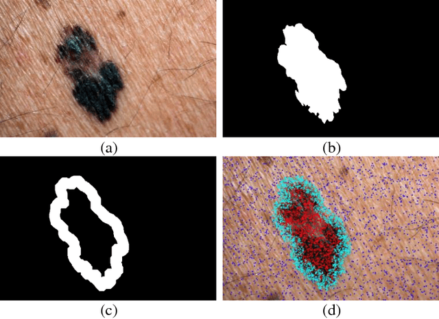
Melanoma is amongst most aggressive types of cancer. However, it is highly curable if detected in its early stages. Prescreening of suspicious moles and lesions for malignancy is of great importance. Detection can be done by images captured by standard cameras, which are more preferable due to low cost and availability. One important step in computerized evaluation of skin lesions is accurate detection of lesion region, i.e. segmentation of an image into two regions as lesion and normal skin. Accurate segmentation can be challenging due to burdens such as illumination variation and low contrast between lesion and healthy skin. In this paper, a method based on deep neural networks is proposed for accurate extraction of a lesion region. The input image is preprocessed and then its patches are fed to a convolutional neural network (CNN). Local texture and global structure of the patches are processed in order to assign pixels to lesion or normal classes. A method for effective selection of training patches is used for more accurate detection of a lesion border. The output segmentation mask is refined by some post processing operations. The experimental results of qualitative and quantitative evaluations demonstrate that our method can outperform other state-of-the-art algorithms exist in the literature.
A Shapley Value Solution to Game Theoretic-based Feature Reduction in False Alarm Detection
Dec 05, 2015False alarm is one of the main concerns in intensive care units and can result in care disruption, sleep deprivation, and insensitivity of care-givers to alarms. Several methods have been proposed to suppress the false alarm rate through improving the quality of physiological signals by filtering, and developing more accurate sensors. However, significant intrinsic correlation among the extracted features limits the performance of most currently available data mining techniques, as they often discard the predictors with low individual impact that may potentially have strong discriminatory power when grouped with others. We propose a model based on coalition game theory that considers the inter-features dependencies in determining the salient predictors in respect to false alarm, which results in improved classification accuracy. The superior performance of this method compared to current methods is shown in simulation results using PhysionNet's MIMIC II database.